RSM Blog: Sports Medicine and Massage Insights
Common Injuries Treated by Deep Tissue Massage
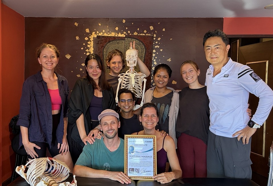
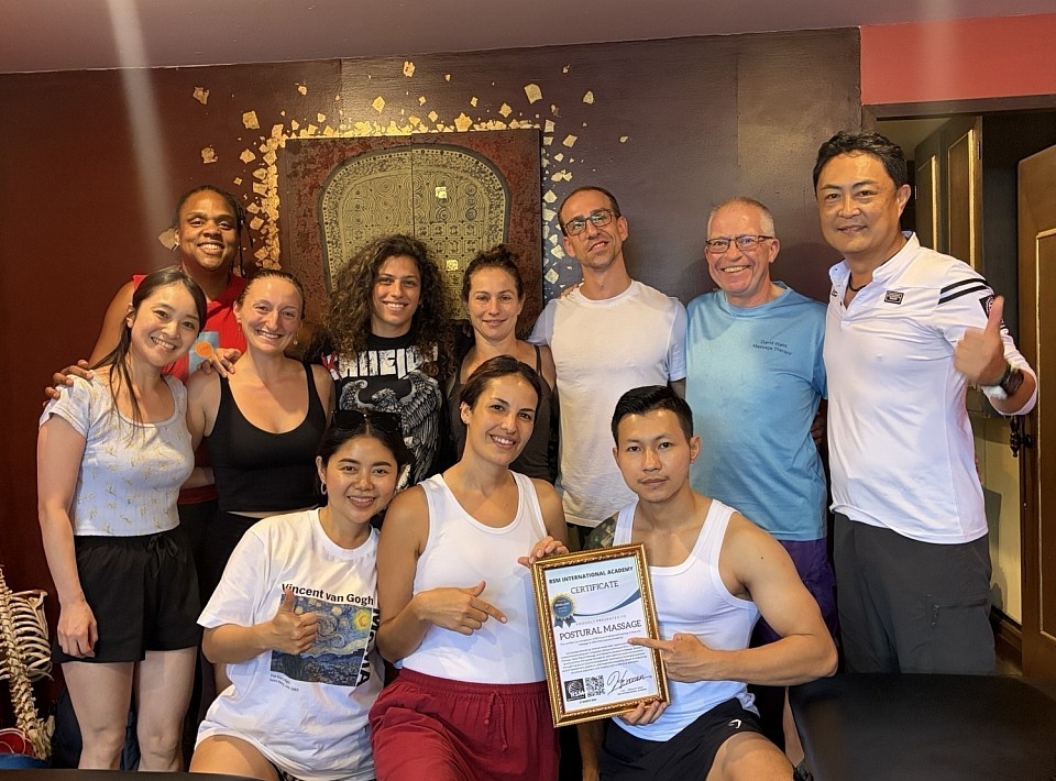
Deep Tissue Masaage For Postural Correction
The human body operates as a kinetic chain where dysfunction in one area inevitably creates compensation in another. At RSM International Academy, we teach students in our Deep Tissue Massage Course that pain is rarely an isolated event; it is a signal of biomechanical failure. When I founded this school in Chiang Mai, my goal was to integrate the precision of sports medicine with the intuitive touch of manual therapy. Effective treatment requires identifying the root cause rather than simply chasing symptoms.
The Mechanics of Deep Tissue Massage
Many clients assume deep tissue massage is defined solely by the amount of pressure applied. This is a misconception. True clinical massage focuses on the specific layers of fascia and muscle that have become adhered. When superficial layers glue to deeper tissues, the sliding mechanism required for healthy movement fails. This friction generates inflammation and restricts range of motion.
Deep pressure applied without anatomical knowledge often triggers protective guarding. Conversely, when a therapist applies sinking pressure that respects the nervous system, they access the deep layers where chronic tension resides. By manually separating stuck fibers, we restore hydration to the fascia. Consequently, the tissue regains elasticity, and the nervous system downregulates the pain signal.
Treating Back Injuries and Pelvic Tension
Low back discomfort is the most prevalent complaint we encounter. However, the source of pain is rarely the lumbar spine itself. In my experience, the lumbar region is often the victim of a tug-of-war between the pelvis and the ribcage.
A primary driver of back injuries is the Quadratus Lumborum (QL). This deep stabilizer connects the hip to the lumbar vertebrae. When a client spends hours sitting, the gluteal muscles become inactive, forcing the QL to work overtime. Over time, the QL becomes hypertonic and short, compressing the lumbar vertebrae.
Standard relaxation massage rarely resolves this because it does not address the depth of the QL. Our students learn to approach the back via the side-lying position to access the anterior edge of the muscle. By releasing this lateral tension, we offload the spine. This mechanical decompression provides longer-lasting relief than simply rubbing the paraspinal muscles.
Sports Injuries and Scar Tissue Remodeling
Athletes subject their bodies to high-velocity loads that frequently result in micro-trauma. Sports injuries such as hamstring strains often heal with disorganized scarring if left untreated.
Deep-tissue massage is critical here because it aligns these repairing fibers. When a muscle tears, the body lays down a patch of dense scar tissue. If this patch remains rigid, the athlete is prone to re-injury because the healthy tissue around it must overwork to compensate for the lack of flexibility.
We treat these injuries massage techniques that apply friction across the grain of the fiber. This transverse friction breaks up adhesions and encourages blood flow. As a result, the tissue heals with greater flexibility. This logic applies to specific conditions like:
- Plantar Fasciitis: Often caused by tight calves pulling on the calcaneus.
- IT Band Syndrome: Frequently a result of tension in the Tensor Fasciae Latae (TFL).
Relieving Chronic Pain and Repetitive Strain
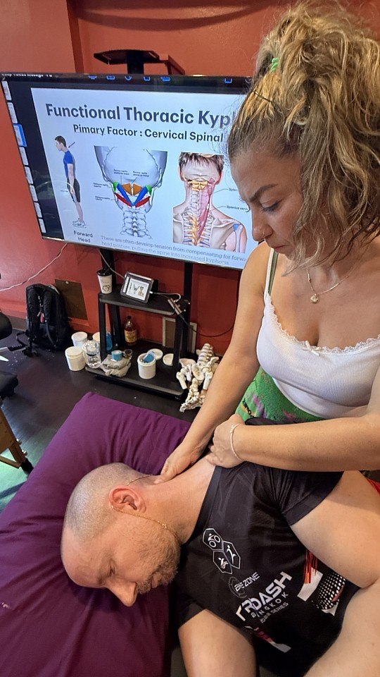
Modern lifestyles force the body into static postures that lead to Repetitive Strain Injuries (RSIs). “Tech Neck” and Upper Crossed Syndrome are common examples where muscle pain is caused by structural imbalance.
In these cases, the chest muscles shorten, pulling the shoulders forward, while the neck muscles lock up to prevent the head from falling. Treating only the neck is futile. To resolve this, we must open the anterior chest wall. Deep tissue work on the Pectoralis Minor allows the shoulders to retract, which naturally relieves the strain on the neck.
Patients often report immediate improvements in numbness or tingling once this proximal tension is resolved. This confirms that the pain in the wrist or arm was actually an entrapment issue higher up the chain.
Systemic Effects of Therapy
Effective therapy extends beyond the physical. Chronic pain disrupts sleep, creating a cycle where the body cannot produce enough growth hormone to repair itself. By breaking the pain cycle through targeted deep-tissue work, we help clients achieve restorative rest. This systemic improvement is one of the most profound effects of manual therapy.
Common injuries, whether from the athletic field or the office, share a root mechanism: the loss of mobility leading to structural overload. By restoring the glide of deep myofascial layers, we allow the body to return to alignment. At RSM, this clinical precision is the standard of care we advocate for in the field of physical medicine.
Essential Resources for Massage Therapy Students: Science & Skills
Faculty Medicine, Chiang Mai University
Foundational Medical Texts for Deep Anatomical Understanding
In my experience as a sports medicine instructor at our massage school here in Thailand, I often see students struggling to locate muscle insertions. This difficulty usually stems from a reliance on the two-dimensional diagrams found in standard massage school curriculums. When a therapist visualizes anatomy as a flat image, their palpation remains superficial. This lack of depth results in ineffective treatment.
To correct this, massage therapy students must invest in high-quality anatomical texts. A resource like Trail Guide to the Body is indispensable because it focuses on palpation paths, teaching you to navigate from a bony landmark to a muscle belly. Another critical text is Gray’s Anatomy for Students, which explains biomechanical relationships. Understanding that the biceps femoris shares an origin with the semitendinosus allows you to treat the entire posterior chain effectively.
I also recommend building a personal library containing reference books on pathology. Knowing the contraindications for conditions like deep vein thrombosis is the primary safety net for any serious practitioner.
Online Tools and Massage Apps for Visual Learners
While books provide intellectual depth, the human body is a dynamic machine. Static images cannot capture how muscle fibers slide during contraction. Consequently, I advise students to supplement their reading with online visualization tools.
Applications like Complete Anatomy allow you to strip away layers of fascia virtually. This digital dissection helps you understand depth, teaching you that treating the piriformis requires sinking through the gluteal mass. Online videos from reputable sources also serve a specific purpose. Watching a cadaver dissection reveals the reality of fascial adhesions ("fuzz"). Seeing the thickness of the thoracolumbar fascia compels you to adopt the deep tissue massage techniques we teach at RSM.
However, be cautious with random social media clips. Always verify what you watch against your anatomy texts.
Why Massage Therapy Journals Matter for Evidence-Based Practice
The field of massage therapy is shifting toward evidence-based medicine. Myths about "flushing lactic acid" are being replaced by physiological facts. To remain respected by other healthcare providers, you must engage with current research.
Accessing a research journal or database like PubMed is crucial. Reading systematic reviews on the efficacy of massage for chronic pain gives you confident talking points based on gate control theory rather than pseudoscience. This habit helps you understand the limitations of bodywork. Recognizing what massage cannot fix is just as important as knowing what it can, ensuring you make correct referrals when necessary.
Investing in Continuing Therapy Education and Mentorship
Graduating from a basic program is only the starting line. Real clinical competence comes from specialized therapy education. At RSM International Academy, we focus on Remedial Massage and Deep Tissue fundamentals because these modalities address the root causes of pain.
Advanced workshops refine your touch. Spending two days focusing solely on the shoulder complex allows you to develop the sensitivity to detect minor adhesions. Mentorship is equally vital. A senior practitioner who critiques your body mechanics can save you from career-ending injury.
When choosing advanced education, look for:
- Instructor Credentials: Teachers should have clinical backgrounds (e.g., Sports Medicine).
- Scientific Basis: Ensure the school curriculum is grounded in anatomy, not unverified theories.
- Hands-on Time: Skill acquisition requires guided practice.
Essential Resources for Career Longevity
Burnout is a high risk for massage therapists. To survive, you must view your body as your most valuable tool and invest in ergonomic resources.
A hydraulic table allows you to adjust height instantly, preserving your lumbar spine. Proper footwear is also non-negotiable; shoes with arch support prevent the plantar fasciitis that leads to compensation patterns in your own body. Finally, peer networks provide psychological support. Joining an association connects you with other massage therapists who understand the demands of the job.
Elevating Your Practice Through Curated Knowledge
The difference between a hobbyist and an expert lies in the resources they utilize. A hobbyist relies on intuition; an expert relies on data and anatomy. At RSM, I emphasize that massage is a cognitive process expressed through the hands. You must think before you touch. Utilize these tools to elevate your standard of care and deliver the specific, science-based benefits your clients deserve.
How to Maintain Proper Body Mechanics

Sports biomechanics and kinetic chain
Understanding Body Mechanics and Kinetic Chains
New students often over-rely on raw muscle power to treat clients. They push with their shoulders and strain their lower backs. This approach inevitably fails. It leads to fatigue and can drastically shorten a therapist’s career. The solution is not strength; it is intelligence. Specifically, we must understand the physics of human movement.
Mechanics acts as the bridge between anatomy and longevity. When I speak of this concept, I refer to coordinating the skeletal, muscular, and nervous systems to maintain balance. If the skeletal structure is stacked correctly, gravity transfers the load through the bones rather than the soft tissue.
However, when alignment breaks, the load shifts. A misaligned joint creates a lever arm that amplifies force on surrounding muscles. Consequently, a small deviation in the hip results in tension elsewhere. This is the kinetic chain. In our massage workshops in Thailand, we teach that you cannot possess a healthy body without respecting these chains.
The Anatomy of Good Posture
Most people view posture as a static position. In reality, it is dynamic. It is the ability to maintain a neutral spine while moving. When the spine is neutral, the natural S-curve absorbs shock efficiently.
Loss of neutrality typically begins in the pelvis. If the pelvis tilts anteriorly, the lumbar spine arches excessively. Conversely, a posterior tilt flattens the lumbar curve, stressing the discs. Moving up the chain, the upper body compensates. We frequently observe the “forward head” position. This forces the trapezius muscles to overwork to support the skull.
To correct this, you must focus on body alignment. We use specific cues:
- Keep the head high, visualizing a string pulling the crown toward the ceiling.
- Retract the chin to align the ears over the shoulders.
- Ensure weight is distributed evenly across the feet.
This “stacking” minimizes muscular effort and allows the skeleton to do the work.
Safe Lifting and Injury Prevention
Whether adjusting a massage table or picking up groceries, the principles of physics remain constant. Improper lifting causes acute lower back issues. The error usually involves bending forward at the waist with straight legs.
When you bend at the waist, you create a long lever arm using your torso. The fulcrum is your lower back. Even a light object becomes heavy due to the torque generated. Avoid this position. The spinal erectors cannot safely lift loads from a stretched position.
Instead, change the mechanics of the movement:
- Approach the object closely to reduce the lever arm.
- Keep your back straight and neutral.
- Descend with your knees bent, pushing your hips back.
- Drive through the heels to stand.
By bending the knees, you utilize the gluteus maximus and quadriceps. Transferring the load to the hips protects the vulnerable spinal muscles. This adjustment is the cornerstone of injury prevention.
The Role of the Body in Massage Therapy
In our curriculum, adhering to proper body mechanics is mandatory. We view the therapist’s body as the primary tool. If the tool is broken, the treatment is ineffective.
When applying deep pressure, a therapist should not push with arm muscles. Pushing requires contraction, which wastes energy. Instead, we teach students to lean. They lock their joints into a safe position and lean their body weight into the client.
This technique utilizes gravity as a limitless source of energy. However, it requires balance. The therapist must maintain a wide stance. The spine remains straight, transmitting force from the legs, through the core, and out through the hands. If a student collapses their chest, the force gets trapped in the shoulder, leading to injury. We correct this by cultivating awareness. If the pressure comes from muscle tension, the mechanics are wrong.
Ergonomics and Proper Body Mechanics for Daily Life
Maintaining health requires vigilance outside the studio. Modern life forces us into sedentary patterns. Sitting in a chair for hours shortens the hip flexors, pulling the pelvis into an anterior tilt when we stand.
To break this cycle, apply care to your environment:
- Adjust monitors to eye level to prevent neck strain.
- Keep feet flat on the floor.
- Position keyboards to keep elbows at 90 degrees.
However, no chair is perfect. The best posture is a changing posture. We recommend moving every 30 minutes to rehydrate fascial tissues. Injury is rarely an accident; it is the result of long-term mechanical negligence.
Why We Prioritize Structural Logic
At RSM International Academy, our philosophy is grounded in science. Hironori Ikeda established this school to elevate standards through physiology, not mysticism. Understanding proper body usage is fundamental.
We analyze the vectors, levers, and loads acting on the system. Whether treating a patient or training a student, the goal is efficiency. By respecting the design of the human skeleton, we ensure longevity.
Implementing proper body mechanics is a discipline. You must catch yourself slouching and correct your form before lifting. Your body is the only vehicle you will ever own. Treat it with the respect a complex machine deserves. Maintain your alignment and move with intention. This is the path to sustainable health.
Essential Ergonomics for Massage Therapists
Sports medicine massage course
The Hidden Ergonomic Hazards in Modern Practice
The career of a manual practitioner is physically demanding. Statistics suggest many graduates leave the industry early due to injury, not from a single accident, but from cumulative micro-trauma. When a provider ignores the mechanics of their own body, connective tissues suffer repetitive strain. This leads to chronic inflammation and instability.
At RSM International Academy, we prioritize the longevity of massage therapists. I often see students in our Trigger Point Therapy Course sacrificing structural integrity for “perfect” technique. This is a fundamental error. Effective therapy requires the provider to operate from a position of mechanical advantage. If the practitioner is unstable, the massage loses efficacy, and the provider risks injury.
The primary culprit is a misunderstanding of force. Many believe pressure comes from upper-body muscle effort. In reality, safe pressure originates from the ground. When the lower body kinetic chain is disconnected, the upper body compensates, loading small joints like the wrist and shoulder which are ill-equipped for high compression.
Understanding the Mechanics of Risk
Ergonomic risk is a calculation of load versus capacity. For instance, leaning over a client with abducted elbows increases torque on the shoulder, forcing the rotator cuff to stabilize aggressively. This narrows the subacromial space, leading to impingement.
To prevent muscle strain and injury, elbows must remain close to the core. This transfers load away from the rotator cuff into the robust latissimus dorsi. Safety is about geometry; a practitioner with perfect leverage can work indefinitely without fatigue.
How Massage Therapists Generate Force Without Strain
The difference between a long career and a short one is the use of gravity versus muscle tension. Muscular effort is metabolically expensive; gravity is free. Proper ergonomics align the skeleton so gravity does the work.
We emphasize “stacking the joints”: aligning wrist, elbow, and shoulder in a vertical line. This bone-on-bone support creates a rigid column to transmit pressure without exhausting the massage therapist. However, stacking requires a lower-body engine. By utilizing a lunge stance, the provider shifts their weight to drive the stroke. The sensation should be one of “falling” into the client, not pushing.
The Role of Proprioception in Working Safely
Proprioception is critical for injury prevention. Practitioners must monitor for “parasitic tension”: unnecessary contractions like clenched jaws or shrugged shoulders. This wastes energy and disrupts the session. By consciously depressing the scapulae, the provider stabilizes the shoulder girdle and reduces neck strain. Correcting these habits reduces the metabolic cost of performing massage therapy.
Optimizing the Massage Table for Biomechanical Efficiency
The massage table height dictates spinal angle and leverage. While “knuckle height” is a common baseline, it is not universal. Deep tissue work often requires a lower table to utilize body weight via a vertical vector. Conversely, detail work requires a higher table to prevent excessive spinal flexion.
If a massage table is too low for detailed tasks, the practitioner must round their back, increasing shear force on discs.
Adjusting for Different Massage Tasks
Hydraulic massage tables are ideal, but if unavailable, the provider must adjust their stance. Widening the stance lowers the center of mass, effectively raising the client’s relative height. Furthermore, distinct massage tasks require distinct working postures. Compression requires vertical stacking; effleurage requires a horizontal lunge.
The workplace must also allow for movement. Cramped rooms force awkward contortions, increasing ergonomic hazards. A spacious room allows the massage therapist to flow around the client, maintaining optimal biomechanics.
Advanced Massage Therapy Mechanics: The Kinetic Chain
Force travels in a wave: from the ground, through the legs, directed by the hips, and into the client. This requires mobile hips and a stable core. If hips are tight, practitioners often compensate by twisting the lumbar spine. However, the lumbar spine is designed for stability, not rotation.
Core Stability and Health
The core protects the spine via Intra-Abdominal Pressure (IAP). Engaging the transverse abdominis supports the lumbar vertebrae during deep compression. Breathing is crucial here; holding breath reduces IAP and spikes blood pressure. Rhythmic breathing maintains stability and promotes a parasympathetic state for both giver and receiver. Maintaining health requires treating the core as safety equipment.
Protecting the Massage Practitioner: Specific Joint Strategies
The thumb and wrist are frequent failure points. The thumb’s CMC joint is designed for grasping, not compression. Relying on thumbs for deep pressure grinds cartilage, leading to osteoarthritis.
We advocate using the elbow and forearm. These robust structures deliver pressure with zero risk to small hand joints. If the thumb must be used, support it with the other hand to distribute force. Additionally, maintain a neutral wrist to spare the median nerve.
Working Postures and Footwear
Poor ergonomics often start at the feet. Elevated heels shift gravity forward, forcing leg muscles to overwork. Zero-drop shoes with wide toe boxes provide a stable base. Furthermore, static postures kill circulation. Constantly shifting weight aids venous return and prevents fatigue.
Integrating Self-Care into Professional Practice
A provider cannot pour from an empty cup. Self-care is a maintenance protocol.
The Recovery Phase
Between clients, the practitioner must reset. Since massage involves flexion, recovery should involve extension, like chest openers or doorframe stretches. Hydration is also paramount to prevent fascial adhesion.
Mental Ergonomics
Physical burnout often follows mental burnout. The emotional labor of treating pain is taxing. Setting boundaries like defined hours and scheduled breaks is an ergonomic strategy. At RSM, we teach that taking on too much leads to fatigue, which compromises biomechanics.
The Hazards of Massage: A Preventable Reality
We must acknowledge that hazards in massage exist, from biomechanical strain to environmental factors. Slippery floors from oil or poor lighting causing eye strain are real risks. A clean, well-lit workplace with good ventilation is an ergonomic necessity to keep the massage therapist alert and safe.
A Commitment to Longevity
The art of massage is sustainable only when the artist is protected. We cannot separate treatment quality from the provider’s health. A practitioner in pain cannot listen to the client’s tissue.
By mastering biomechanics and utilizing gravity, the provider transforms labor into a rhythmic dance. This is the core philosophy at RSM International Academy. Through intelligent practice, appropriate equipment, and self-care, the ergonomic risks are mitigated. We aim to build massage therapists designed to last, ensuring their hands can continue to heal others for decades.
The Massage Therapy Code of Ethics and Professional Standards
Sports medicine massage school
Defining the Core of Our Practice
I teach students in our Remedial Massage Course that technical skill without a moral compass easily results in clinical failure. We study anatomy to understand movement and pathology to identify dysfunction. However, we study ethics to ensure that our application of this knowledge remains safe.
When a client enters our clinic, they are often in pain. Pain alters the nervous system, increasing sympathetic arousal and vulnerability. If a professional acts without integrity, that vulnerability transforms into guarding. Muscle tension increases, and the therapeutic window closes. Therefore, adhering to a strict framework of conduct is not merely a legal requirement; it is a physiological necessity for healing.
I view the relationship between a therapist and a client as a contract of trust. This contract allows us access to the body’s soft tissues. In return, we must guarantee safety through a deep understanding of boundaries and confidentiality. Without these pillars, even the most advanced massage therapy techniques will fail to produce lasting results.
Why a Strict Code of Ethics Matters
Many people view ethics as a list of restrictions. I teach it as a set of operating parameters that optimize clinical outcomes. Just as a surgeon follows sterile protocols, a massage therapist follows ethical protocols to prevent harm.
When a client trusts their therapist, their parasympathetic nervous system engages. Heart rate drops, and muscle tone decreases. This state facilitates deep bodywork. Conversely, uncertainty about a therapist’s intentions triggers the sympathetic nervous system. Cortisol levels rise, and muscles tighten defensively. As a result, the manual pressure we apply meets resistance rather than acceptance.
A rigid code of ethics balances the inherent power dynamic in the treatment room. It ensures that we use our position solely for the client’s benefit. At RSM, our commitment to excellence aligns with global benchmarks. We look to standards like the AMTA code or ABMP code to guide our principles. These organizations provide a framework that elevates massage from a service to a healthcare discipline.
Ensuring Client Safety and Trust
Safety is the primary directive of any healthcare intervention. In massage therapy, safety encompasses physical, emotional, and informational security. If any of these are compromised, the therapy ceases to be effective.
True safety begins with informed consent. Before I touch a client, I explain the plan. I detail which muscles we will target and why. This clarity removes fear. When the client knows what to expect, they remain in control and can relax. If a therapist skips this step, the client remains on high alert, rendering the treatment ineffective.
Information security is equally critical. Clients share sensitive health history and personal stressors with us. If a therapist breaches this confidentiality, trust evaporates. We treat client records with the same rigor as a hospital, ensuring that privacy is paramount.
Professional Conduct in a Clinical Setting
Professional conduct is the visible manifestation of our ethical framework. It is how we dress, speak, and manage the environment. Clients judge a therapist’s competence before the session begins. They assess the cleanliness of the room and the clarity of communication.
Pathogens do not respect intentions; they only respect hygiene. Because we work in close contact with skin, a failure in hygiene leads to cross-contamination. Therefore, we enforce strict sanitation policies at RSM. We also consider personal hygiene. Strong perfumes can trigger allergic reactions or headaches, forcing the client to endure the session rather than enjoy it. We aim for neutrality – neutral scent and neutral demeanor – to create a blank canvas for the therapy.
One of the most frequent violations involves the scope of practice. Massage therapists are not doctors. We do not diagnose. We assess soft tissue function. When a therapist steps outside their lane, they endanger the client. Telling a client they have a “slipped disc” based on palpation creates fear (nocebo effect), which increases pain perception. Instead, we refer them to specialists, respecting our limits and prioritizing their long-term health.
Navigating Ethical Dilemmas in Bodywork
Therapists inevitably face gray areas. These dilemmas require critical thinking. A common issue is the “dual relationship,” where a therapist and client have a connection outside the treatment room. This blurring of lines complicates the clinical dynamic and can suppress honest feedback. To manage this, we must establish clear separation, focusing strictly on anatomy and the treatment plan during the session.
We also navigate transference and counter-transference. Sometimes, touch is misinterpreted as affection. If I notice a client becoming overly attached, I must re-establish boundaries immediately. I might use more clinical language or adjust the draping. If the behavior continues, I must terminate the services. The integrity of our ethical practices depends on this discipline.
High Standards in Massage Education
The quality of a therapist is directly proportional to their education. At our school, we simulate ethical challenges in the classroom. We do not wait for students to enter the workforce to learn these lessons.
Draping, for example, is not just about modesty; it is about security. Proper draping creates a clear boundary, allowing the client to disassociate from their vulnerability and focus on the treatment. We teach secure tucking and minimal exposure. This precision distinguishes a professional from an amateur.
Ethics also extends to financial integrity. We reject the “hard sell.” We do not promise miracle cures or up-sell unnecessary add-ons. We treat the client’s resources with the same respect we treat their body. In a landscape where massage is often commodified, operating with transparency builds a reputation of reliability.
Elevating Client Care Through Science
I established RSM to bridge the gap between traditional massage and modern sports medicine. Science and ethics are inextricably linked. Using outdated techniques is an ethical failing because it wastes the client’s time.
We rely on ethical standards grounded in evidence. For instance, we know that aggressive foam rolling on the Iliotibial Band is often ineffective mechanically. The tension usually originates in the Tensor Fasciae Latae (TFL). Therefore, treating the TFL is the ethical choice because it is the effective choice. By prioritizing efficacy, we honor the client’s trust.
This approach requires rigorous assessment. Without assessment, we are guessing. If I treat shoulder pain without checking for a rotator cuff tear, I risk aggravating the injury. The assessment dictates the treatment. This logical sequence protects the client and validates our massage code.
The Foundation of Excellence
I tell my students that their hands can only take them so far. It is their character that sustains their career. A professional who operates with integrity builds a loyal following.
At RSM International Academy, we teach responsibility. We teach that the clients on our tables are people with complex nervous systems. By adhering to rigorous ethical codes, we create a safe space where tissues can heal and where the profession of massage therapy can thrive. Science informs our hands, but ethics guides our hearts. Together, they create the complete therapist.
Scientific Perspectives on the Benefits of Shiatsu Massage Techniques
Deep shiatsu massage course
The Physiological Mechanisms of Shiatsu Massage
At RSM we approach every modality through the lens of sports medicine, but often encounter the misconception that Eastern modalities rely solely on esoteric concepts. While the traditional concept of Qi is historically significant, the efficacy of shiatsu massage is grounded in tangible anatomy and physiology.
When we apply perpendicular static pressure, we engage a distinct physiological chain reaction. Unlike dynamic oil massage, shiatsu utilizes sustained compression. This compression temporarily reduces local blood flow (ischemia). Upon release, the body responds with a rush of fresh, oxygenated blood (hyperemia) to the area. This results in improved circulation, actively clearing metabolic waste like lactic acid. Therefore, the massage techniques we teach in our Shiatsu Massage Course are mechanical interventions designed to restore homeostasis.
Correcting Body Alignment with Static Pressure
Structural misalignment often stems from chronic muscle shortening. A shortened muscle pulls on its tendon, which in turn alters the position of the bone it attaches to. This creates a cascade of imbalance throughout the entire body.
Shiatsu massage addresses this biomechanical issue by applying deep, static pressure to the belly of a hypertonic muscle. This inhibits the muscle spindles; sensory organs that detect stretch. As a result, muscle tone decreases, and fiber length is restored. For example, by releasing a tight psoas muscle, a skilled practitioner can reduce the pull on the lumbar vertebrae, effectively treating the root cause of lower back pain rather than just masking symptoms.
How Shiatsu Regulates Autonomic Function
Modern lifestyles keep the sympathetic nervous system (fight or flight) in overdrive, leading to elevated cortisol and delayed tissue repair. As a sports medicine specialist, I view this stress state as a barrier to healing.
Massage therapy acts as a modulator. The rhythmic and static nature of shiatsu mimics the body’s natural biorhythms. The brain interprets this sensory input as a safety signal, switching the body into a parasympathetic (rest and digest) state. This hormonal shift is critical; without it, the body cannot effectively repair micro-trauma in muscle fibers or achieve deep recovery.
Managing Health and Pain Perception
Pain is complex, acting as an output of the brain based on perceived threat. regarding pain, shiatsu utilizes the “Gate Control Theory” to manage discomfort and improve health.
When a therapist applies firm, non-painful pressure, large-diameter nerve fibers are activated. These fibers transmit signals to the spinal cord faster than the small-diameter fibers that transmit pain. Therefore, the pressure signal “closes the gate” to the pain signal. By treating trigger points in this manner, we disrupt the pain-spasm-pain cycle. The treatment resets the neuromuscular junction, allowing the muscle to return to a resting state.
Enhancing Mental Wellbeing Through Massage
Physical recovery cannot be separated from mental state. The two are intrinsically linked via the psychosomatic loop. To support mental well-being, shiatsu targets this connection directly.
Massage breaks this loop. By reducing physical tension in the neck and shoulders, we lower the sensory “noise” reaching the brain. This is vital for sleep. Sleep is the only time the body releases growth hormone in significant quantities for tissue repair. By lowering cortisol and inducing relaxation, shiatsu prepares the neurochemistry for deep, restorative sleep. Therefore, we view wellbeing as a quantifiable clinical outcome.
Integrating Treatments into Sports Medicine
At RSM, we do not view shiatsu as alternative therapy; we integrate it as a vital tool for athletic recovery. Athletes require optimal flexibility and range of motion. However, overuse often causes connective tissue (fascia) to become dehydrated and adhered.
The shearing and compressive forces used in shiatsu bodywork help rehydrate fascia and break down adhesions. This improves the glide between muscle layers. Specifically, by treating “myofascial chains” such as the posterior line from foot to head, we address global tension patterns. Regarding local circulation, shiatsu ensures that oxygen reaches these deep tissues, allowing the athlete to perform at peak efficiency.
The Importance of Professional Training
The benefits discussed above – autonomic regulation, structural correction, and pain management – rely heavily on the skill of the therapist. Random pressure provides random results. To achieve clinical outcomes, one must understand anatomy, pathology, and biomechanics.
At RSM International Academy, we teach students to palpate with precision, distinguishing between hypertonic muscle and fibrotic tendon. It is this dedication to science that elevates simple massage therapy into a medical-grade intervention.
Practical Application: Hand Self-Shiatsu
While professional treatment is irreplaceable, I often advise clients to perform maintenance between sessions. One simple technique is hand self-shiatsu.
The thenar eminence (the thumb base) accumulates tension from modern device use. By using the opposite thumb to apply deep, static pressure into this muscle pad for 3-5 seconds, you can release tension that refers up the arm. This simple act can temporarily improve circulation and reduce local fatigue.
How Sports Massage Improves Performance: The Physiological Mechanisms
Sports massage course
The Role of Sports Massage in Recovery
When we analyze how sports massage improves performance, we must first look at the mechanics of recovery. Intense physical exertion creates micro-trauma in the muscle fibers and generates metabolic waste. While the body naturally clears this waste, the process relies heavily on muscle contraction to pump venous blood and lymph back to the heart.
In RSM's Sports Massage Course, we teach that sports massage acts as a mechanical assist to this system. By manipulating soft tissue, we create external pressure gradients that force fluid out of interstitial spaces and into the vascular network. This accelerates the delivery of oxygen and nutrients required for repair. Consequently, the downtime between training sessions decreases. This allows athletes to maintain higher volumes of work without succumbing to fatigue.
Reducing Muscle Soreness and Inflammation
One of the most immediate barriers to high-level output is pain. Muscle soreness, particularly Delayed Onset Muscle Soreness (DOMS), alters movement mechanics. When an athlete moves to avoid pain, they develop compensatory patterns that leak energy. It is vital to understand that this soreness is largely driven by inflammatory markers.
Research suggests that targeted therapy modulates this inflammatory response. By reducing inflammation, we prevent the “guarding” reflex where the nervous system tightens tissue to protect a painful area. At RSM, we use specific pressure to interrupt pain signals, a concept known as the Gate Control Theory. This lowers the perception of pain and allows for normal functional movement to return faster.
Optimizing Jump Performance Through Flexibility
Power is the product of force and velocity. In sports requiring explosive movement, restricted tissue is a major liability. We see this clearly when analyzing jump performance. The posterior chain must lengthen rapidly before contracting. If the hamstrings or calves are hypertonic, they limit the elastic energy storage required for a powerful jump.
Regular sports massage normalizes the length-tension relationship of these muscles. We mobilize the fascial layers to ensure they glide efficiently. When internal friction is reduced, the tendon can load effectively like a spring. This compliance allows the athlete to express their full power potential. By restoring ankle range of motion through deep calf work, we also allow for a deeper loading phase, which directly correlates to vertical height.
Essential Massage Techniques for Athletes
Clinical application requires a precise strategy. The massage techniques used must match the physiological goal of the athlete’s current cycle.
- Effleurage: Long, gliding strokes used to warm the tissue and assist lymphatic drainage.
- Petrissage: Kneading and lifting the muscle belly to separate fibers and break down adhesions.
- Friction: Concentrated pressure to remodel scar tissue in tendons.
Understanding when to apply these is key. For recovery, we use slower strokes to recruit the parasympathetic nervous system. Conversely, pre-event work utilizes rapid percussion to excite the nervous system. A massage therapist must be able to read the client’s autonomic state to deliver the correct stimulus.
Why Athletic Individuals Need Maintenance
There is a distinction between treating an injury and optimizing a system. Many athletic individuals wait until structural failure occurs before seeking help. This reactive approach is inefficient. Maintenance therapy serves as a diagnostic checkpoint.
By assessing tissue quality regularly, we can detect tightness in areas like the hip flexors or calves before they cause a tear. This proactive management prevents cumulative load from reaching the threshold of injury. For professional athletes, this consistency is often the difference between a long career and early retirement.
The Role of the Massage Therapist
The relationship between the massage therapist and the coach should be collaborative. We periodize treatments just as coaches periodize lifting. During high-volume build phases, we focus on systemic flushing and deep tissue pliability to manage fatigue. As competition approaches, we shift to mobilization. We avoid aggressive work that might lower muscle tone too much, as a certain amount of tension is required for reactivity.
Integration with Physical Therapy
Finally, it is crucial to integrate manual work with physical therapy. While PT focuses on corrective exercise, massage provides the mobility required to execute those exercises. If a hip capsule is too tight, a client cannot squat correctly. By releasing the restriction manually, we create a window of opportunity for strengthening.
This synergy is the key to improving performance. By prioritizing muscle recovery, neurological balance, and mechanical efficiency, we ensure that every ounce of effort in the gym translates to success on the field. This clinical precision is the standard we uphold at RSM.
Managing Client Expectations in Massage for Better Clinical Outcomes
Practical sports medicine massage techniques
At RSM we recognize the importance of preparing students in our Remedial Massage Course to become professional therapists. One point to be realized is that learning advanced anatomy and acquiring technical skills alone does not ensure a thriving practice. Even the most gifted therapist will struggle if they cannot navigate the psychological side of the treatment room. Specifically, success depends on the ability to control the narrative regarding recovery.
Clients arrive at an appointment with a pre-existing belief system. They often view pain as the problem rather than a signal. Consequently, they expect the pain to vanish within a single session. If we allow this belief to persist, we set ourselves up for failure. Tissue healing follows a biological timeline. Inflammation must subside, and collagen must remodel. My role is to teach you how to align the client’s mind with their physiology.
Establishing Clear Communication During Assessment
The foundation of success lies in the intake. This is where you gather information and establish clinical authority. Too many therapists rush to get the client on the table. This is a mistake.
Consider a client with lateral knee pain. They point to the knee and ask for deep pressure. A novice complies. An expert investigates. We know that lateral knee pain often stems from pelvic anterior tilt, which shortens the Tensor Fasciae Latae (TFL). A tight TFL pulls on the Iliotibial Band, creating friction at the knee.
When you explain this chain, Pelvis → TFL → IT Band → Knee, you shift their focus. They stop expecting magic at the knee and start understanding the necessity of treating the hip. This intellectual alignment is the first step in managing what they expect from the therapy.
The Psychology of Pain and Recovery in Massage
Clients often gauge the success of a massage solely by pain reduction. However, breaking down adhesions releases inflammatory byproducts. This causes temporary soreness (DOMS).
If you do not warn a client about potential soreness, they will interpret it as an injury. Conversely, if you predict the soreness, they view it as a sign of progress. I teach my students a simple script: identify the restriction, explain that releasing it causes inflammation, and frame the resulting soreness as the healing response. By predicting the outcome, you build trust.
Realistic Expectations for Chronic Conditions
Acute injuries have clear healing curves. Chronic pathologies do not. A client with Upper Cross Syndrome from years of office work cannot be “fixed” in one hour. They spend 40 hours a week creating the problem. One hour of therapy cannot mathematically undo that damage; it can only mitigate it.
We use this logic to propose a plan. We do not sell single sessions; we propose a course of treatment. This structure creates realistic expectations. The client stops looking for a miracle cure and starts looking for incremental progress.
Client Feedback and the “No Pain, No Gain” Myth
A pervasive myth exists that massage must be excruciating to work. You will encounter clients who demand maximum pressure, believing intensity equals value. It is your duty to correct this client’s expectation.
I explain the anatomy of the muscle spindle. If I force an elbow into a tight muscle too quickly, the spindle fires, triggering a stretch reflex. The muscle contracts to protect itself. In contrast, if I sink in slowly, the spindle remains quiet. When you explain this biological fact, clients usually relent. They accept a slower, more calculated pace.
We also distinguish between “pain that hurts” and “pain that heals.” We ask, “Is this pressure sustainable?” If they tense up, the sympathetic nervous system activates, fighting our work. A client-centered approach uses feedback to ensure the parasympathetic system remains dominant, allowing for true tissue release.
Elevating Client Expectations Through Clinical Excellence
Client satisfaction is rarely about the technique alone. It is about the alignment between what was promised and what was delivered. Education bridges this gap.
At RSM, we believe that leadership in the treatment room means guiding the client through the process of recovery. It means using anatomy to explain pathology and logic to set goals. When you master this, you stop chasing impossible results. You attract a clientele that values skill over servitude. This is the definition of clinical therapy, and it is the standard we uphold.
FAIR test at Approximately 60 Degrees of Hip Flexion as the Most Revealing Diagnostic Position
Sports massage course chiang mai, thailand
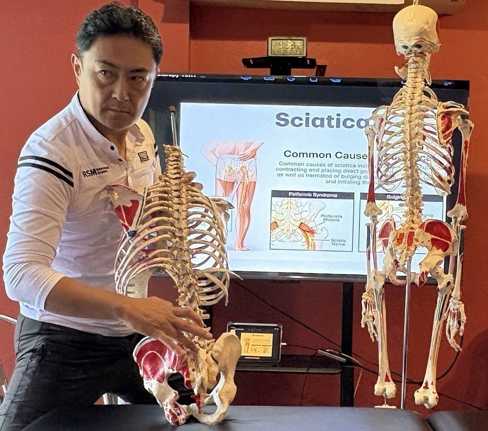
For many years I have evaluated clients whose hip or buttock pain sharply increases during the FAIR position. FAIR refers to flexion, adduction and internal rotation of the hip, and the position becomes most diagnostically accurate when the hip is flexed to approximately sixty degrees. At this angle the deep-gluteal interval tightens in a very specific way. The piriformis begins to shift its rotational function, the obturator complex becomes tensioned, and the sciatic corridor narrows just enough to expose underlying dysfunction that is often invisible in neutral hip positions. This sixty-degree window is where countless clients—athletes, office workers, older adults, people recovering from injuries and individuals who simply sit too long—show their clearest symptoms.
Across decades of hands-on work, one observation has repeated itself with remarkable consistency. People who develop lateral-hip or trochanteric pain often walk with a pronounced lateral pelvic shift. It is especially common in heavier individuals and in many women who demonstrate a side-to-side pelvic drop with each step. The mechanism is simple when you see it enough times. Excess pelvic shift increases compression over the gluteus medius and minimus tendons and the trochanteric bursa. FAIR—because it exaggerates adduction and internal rotation—magnifies the same forces that irritate these tissues during walking. For these clients the FAIR test is not a piriformis problem at all. It is a reflection of chronic lateral-hip overload that has been silently developing through years of compensatory gait.
Deep buttock pain during FAIR often reveals irritation within the sciatic corridor rather than an isolated piriformis syndrome. Many clients spend decades sitting with a habitual pelvic rotation, a leg crossed over the other or a consistently flexed lumbar posture. These patterns bind the deep gluteal fascia, limit capsular movement and create a narrow channel in which the sciatic nerve becomes vulnerable. FAIR does not create this vulnerability. It merely exposes it. I have seen this pattern in athletes and in people who have never trained a day in their lives.
There is also a group whose FAIR-induced pain appears not in the buttock but in the medial thigh. This clinical picture is almost always tied to the obturator nerve or to tension within the fascial envelope of the adductor complex. Hip adduction and internal rotation load this region more than most people realize. Many clients who sit long hours, who have old groin strains or who lack genuine hip-rotation capacity experience a distinct medial-thigh pain during FAIR. In these cases the problem is neither piriformis nor sciatic. It is a diagnostic doorway into a deeper issue of obturator-related irritation that is frequently misclassified in standard practice.
Another consistent pattern emerges in clients with lateral hip pain and tenderness around the greater trochanter. These individuals may sleep predominantly on one side, collapse their hip during walking, or rely on compensatory tension in the iliotibial band to stabilize a weak pelvic structure. When placed in FAIR, the lateral hip becomes compressed under conditions identical to those experienced during daily walking. What appears to be a positive piriformis test is, in reality, a clear sign of greater-trochanteric overload and gluteal-tendon sensitization.
I have encountered a different subset of clients in whom FAIR provokes deep posterior-hip discomfort without sciatic characteristics. This arises when the deep external rotators—obturator internus, obturator externus, the gemelli, or quadratus femoris—lose their stabilizing function. These muscles quietly hold the femoral head inside the acetabulum during ordinary tasks such as climbing stairs, rising from a chair or simply turning in bed. When they fail, the FAIR position reveals their weakness instantly. The discomfort is unmistakable for anyone who has palpated these structures as often as I have.
Finally、there are individuals who feel a sharp or aching sensation near the ischial tuberosity during FAIR testing. This region hosts not only the proximal hamstring origin but also the pathways of the sciatic nerve and the posterior femoral cutaneous nerve. People who sit on firm surfaces for long periods, long-distance drivers, and those with old hamstring injuries often fall into this category. The FAIR position tensions the posterior-thigh structures far more than it loads the piriformis, and recognizing this distinction is essential for accurate diagnosis.
After years of observing thousands of bodies, the conclusion is clear. FAIR-related hip pain is not a single condition. It is a cluster of different anatomical patterns expressed through one provocative position. Deep buttock pain aligns with the sciatic corridor. Medial-thigh discomfort reveals obturator or adductor fascial involvement. Lateral-hip pain exposes gluteal-tendon compression and trochanteric overload. Posterior-hip tightness arises from deep-rotator dysfunction. Pain near the ischial tuberosity reflects irritation of the posterior femoral cutaneous nerve or proximal-hamstring tissues. Each pattern has a unique mechanical origin and demands a specific therapeutic strategy.
At RSM International Academy, these distinctions are not theoretical. They are the foundation of how we teach manual therapy, movement interpretation and corrective intervention. Students learn to read the FAIR position not as a simple piriformis test but as a diagnostic system that reveals the deeper organization— or disorganization—of the hip. Whether through Sports Massage, Trigger Point Therapy, Deep Tissue Massage, Remedial Massage or Dynamic Myofascial Release, the goal is always the same. Understand the pattern. Touch the truth beneath the surface. Intervene with precision, not assumption.
- Hironori Ikeda, MSc sports medicine
Neurodynamics & Sports Biomechanics Specialist
References
1) Benson E et al. Deep gluteal syndrome and posterior hip pain. Clinical Orthopaedics
2) Bradshaw C and McCrory P. Obturator nerve neuropathy and groin pain patterns. Sports Medicine.
Tennis Elbow and Golfer’s Elbow as Failures of Wrist Centrifugal Adaptation and Shoulder Kinetic Chain Function
Sports massage course students
Lateral and medial epicondylitis have traditionally been described as local overuse injuries involving the wrist extensors and flexors. From a modern sports medicine and biomechanical perspective however these conditions are better understood as the result of two interacting problems. The first is the inability of the wrist to adapt to rapidly increasing centrifugal force at impact. The second is a breakdown of the kinetic chain at the glenohumeral joint which forces the forearm to compensate through excessive pronation and supination.
During a tennis stroke a golf swing or any striking motion the racket club or bat generates a marked increase in centrifugal force from impact into the follow through. A healthy wrist responds to this force by allowing a brief delayed motion that creates functional spacing between the carpal bones and the radius and ulna within the wrist joint. This spacing allows traction forces to be dissipated across the entire wrist complex rather than transmitted directly into the elbow. The carpus and distal forearm effectively work as a shock absorber that protects the epicondylar tendon origins from excessive eccentric overload.
When an athlete stiffens the wrist with excessive muscular co contraction this protective spacing is lost. The wrist can no longer absorb the centrifugal pull of the racket or club and the resulting force is transferred almost directly to the forearm muscles. The wrist extensors and flexors are then subjected to rapid eccentric loading at the moment of impact. Over time this leads to microtrauma and degenerative change at the lateral or medial epicondyle which is the classic presentation of tennis elbow and golfer’s elbow. Biomechanical studies have repeatedly shown that rapid eccentric overload of the wrist extensors during impact is a key mechanism in the development of lateral epicondylitis.
The picture becomes more complex when we consider the glenohumeral joint. In efficient overhead and striking mechanics the shoulder provides a substantial portion of the rotational motion required for stroke production through well coordinated internal and external rotation. When glenohumeral internal rotation is limited a condition often referred to as GIRD the athlete may attempt to square the racket face or clubface by increasing forearm pronation. Conversely when external rotation is insufficient the athlete may rely excessively on supination. In both cases shoulder rotation that should occur at the GH joint is shifted distally into the forearm and elbow.
This compensatory strategy disrupts the kinetic chain. Instead of the shoulder trunk and lower body sharing the load the elbow is forced to manage both the rotational demands of the stroke and the high eccentric forces generated at impact. The elbow becomes a bottleneck in the energy flow. Several kinetic chain studies have shown that altered shoulder mechanics and deficits in total rotational motion are associated with an increased risk of elbow injury in throwing and racket sport athletes.
In this framework tennis elbow and golfer’s elbow are not simply local tendon pathologies. They are manifestations of a global mechanical problem in which the wrist fails to create adequate space and shock absorption under centrifugal load and the shoulder fails to contribute its share of rotational motion. The forearm responds with excessive pronation or supination and the epicondylar structures are exposed to repeated eccentric stress.
Effective prevention and rehabilitation therefore require more than localized treatment of the elbow. They must address the ability of the wrist to move freely and absorb force and they must restore glenohumeral internal and external rotation so that the kinetic chain from shoulder to wrist can function as a coordinated system. Athletes who develop a supple wrist and an efficient shoulder driven kinetic chain can dissipate impact forces far more effectively and significantly reduce the mechanical burden placed on the elbow.
At RSM International Academy these biomechanical principles form the foundation of our advanced professional training curriculum. Our Sports Massage and Remedial Massage programs provide an in depth understanding of kinetic chain mechanics for overhead athletes including glenohumeral internal and external rotation forearm pronation and supination and the wrist’s shock absorption mechanism under centrifugal load.
In our Dynamic Myofascial Release course students study fascial dynamics joint capsule mobilization and upper extremity kinetic chain integration with exceptional depth and precision. These skills represent essential competencies for elite sports trainers and performance clinicians. The curriculum is specifically designed to prepare practitioners to evaluate and correct dysfunctional movement patterns in athletes at every level ensuring they gain the advanced skill set required in modern sports medicine.
- Hironori Ikeda, MSc sports medicine
Neurodynamics & Sports Biomechanics Specialist
Reference
1) De Smedt T et al. Lateral epicondylitis in tennis: update on aetiology diagnosis and treatment. British Journal of Sports Medicine.
2) Riek S et al. A simulation of muscle force and internal kinematics of the forearm during backhand stroke in tennis. Journal of Biomechanics.
Inferior Cluneal Nerve Pain in Pregnancy – A Practical and Biomechanical Perspective from Sports Medicine
Sciatica pain treatment
Pregnancy produces a remarkable sequence of biomechanical changes that reshape the spine, pelvis and surrounding soft tissues. In clinical work, and even in observing people in everyday life, it is clear how rapidly posture begins to shift as the abdomen grows. The deep stabilizing system — the transversus abdominis, diaphragm, pelvic floor and obliques — slowly loses mechanical advantage. When this support decreases, the lumbar spine naturally increases its lordosis, and the pelvis slides into a stronger anterior tilt. With this shift, the sacrum almost inevitably moves into nutation, tilting forward and increasing pressure at the back of the pelvis.
These adaptations are not pathological; they are part of human design. But when they combine with relaxin-driven ligament laxity, the sacroiliac joint becomes more mobile than usual. This allows small shear forces to appear around the sacrum—forces that are normally well-contained. Over many weeks, these micro-movements affect the tissues at the gluteal fold, exactly where the inferior cluneal nerve travels beneath the lower edge of the gluteus maximus.
I often see how gait changes during pregnancy: a slightly wider stance, external rotation of the hips, and a subconscious attempt to balance the shifting center of gravity. These adjustments place extra recruitment demands on the gluteus maximus and the deep external rotators. When these muscles tighten, especially the piriformis, they transmit tension into the area where the inferior cluneal nerve branches from the posterior femoral cutaneous nerve. This is why many pregnant women feel a burning or sharp pain at the lower buttock — sometimes radiating gently into the upper hamstring. It looks like sciatica to the untrained eye, but the pattern, when evaluated properly, fits cluneal nerve irritation with surprising accuracy.
Sitting becomes another source of stress. The gluteal fold must bear more body weight, and with sacral nutation plus pelvic tilt, the available space around the nerve decreases. This is why symptoms increase after long periods of sitting on firm surfaces. It is not random pain — it is physics, weight distribution, and altered anatomy working together.
This entire process forms a predictable chain reaction: abdominal expansion reduces deep core support; the lumbar spine compensates; the pelvis tilts; the sacrum nutates; ligaments soften; shear forces rise; and hip external rotators tighten. When put together, they create a perfect environment for inferior cluneal nerve compression.
For many families, especially where cultural boundaries make it difficult for male therapists to treat female clients, partners often feel helpless. Yet when the mechanism is explained — not as a mysterious “pregnancy pain,” but as a clear biomechanical sequence — husbands and partners suddenly understand what is happening. With basic knowledge, they can help their wives with simple soft-tissue work, pelvic unloading positions, or small posture adjustments that meaningfully reduce pressure on the nerve. This practical understanding often has more impact than people expect.
From a sports medicine standpoint, the condition responds extremely well to conservative approaches. Gentle soft-tissue release at the lower gluteal border, mild sacral decompression positions, controlled breathing to re-engage intra-abdominal pressure, and easy posterior pelvic tilt exercises can all reduce stress on the nerve. These interventions work because they directly counter the mechanical pathway that created the problem. Misdiagnosis, however, leads to treatments that often worsen symptoms — particularly aggressive stretching or unnecessary lumbar-focused therapy.
Scientific literature strongly supports this understanding. Vleeming and colleagues describe how sacroiliac joint instability increases during pregnancy, aligning closely with the mechanical explanation above. Kuniya’s anatomical study maps the cluneal nerves with precise detail and shows how subtle shifts in sacral angle can irritate these nerves at identifiable entrapment points. These studies have consistently matched what I see in real people: when the pelvis changes, the nerves respond.
Pregnancy-related inferior cluneal nerve pain is not an accident. It is a predictable outcome of how the human body adapts to carrying new life. When explained clearly, clinicians, partners and mothers themselves can recognize the early signs and manage the condition with confidence and clarity. Understanding turns fear into calm, and simple strategies can prevent weeks of unnecessary suffering.
- Hironori Ikeda, MSc Sports Medicine
Neurodynamics & Sports Biomechanics Specialist
References
1) Vleeming A, et al. “European Guidelines for the Diagnosis and Treatment of Pelvic Girdle Pain.” European Spine Journal, 2008
2) Kuniya H, et al. “Anatomical Study of the Cluneal Nerves and Their Entrapment Sites.” Pain Physician, 2013.
Understanding Ankle Dorsiflexion Limitation and Its Impact on Lumbar and Hip Compensation in Sports Movement
Sports biomechanics study
In many athletes and general clients I’ve worked with over the years, one of the most consistent patterns I observe is how a simple restriction in ankle dorsiflexion can quietly influence the entire kinetic chain. It rarely stays at the ankle. The moment dorsiflexion becomes limited—whether from muscular tightness, joint restriction, or deep fascial tension—the body tries to find a way around it. And the compensation almost always travels upward into the knee, the hip, and eventually the lumbar spine. Once you see this pattern enough times in real people, it becomes impossible to ignore.
When the ankle cannot dorsiflex properly, the knee loses its ability to translate forward in a natural way. This forces the hip to take on more flexion than intended, and in many athletic positions—especially the so-called “power position”—the lumbar spine begins to extend excessively to maintain balance. This is one of the hidden pathways by which dorsiflexion limitation contributes to low back discomfort. It’s not a dramatic shift; it’s subtle. But repetition during walking, training, lifting, or sport compounds the stress. The more limited the ankle is, the more the lumbar spine and hip must compensate.
One of the primary reasons dorsiflexion becomes restricted lies in the posterior compartment of the lower leg—particularly the gastrocnemius and soleus. Shortening, chronic tightness, or active trigger points in these muscles reduce the available tibial translation over the talus. But the deeper contributors are just as important: the tibialis posterior, flexor hallucis longus, and flexor digitorum longus often create a kind of “deep compartment stiffness” that many clinicians overlook. These deeper muscles don’t scream for attention, but they significantly alter ankle mobility when they tighten.
Just as critical is the movement of the talus itself. Proper dorsiflexion depends on adequate posterior glide of the talus. When this glide becomes restricted—whether due to joint capsule stiffness, retinaculum tension, local swelling, or even decreased sliding of the fat pad—the ankle simply cannot express its full range. Without this posterior movement, the tibia is forced to compensate, and the chain reaction quickly spreads upward. In my experience, once talar mobility is restored, many seemingly unrelated movement problems begin to improve almost immediately.
Athletes often feel this during squatting, lunging, or deceleration drills. With limited dorsiflexion, they shift their weight backward, externally rotate at the hip to “open space,” or overextend the lumbar spine to stay upright. These are not conscious choices; they are automatic compensation strategies the body uses to keep the movement going. But these same patterns, repeated daily, become a source of stress for the lumbopelvic system.
Addressing dorsiflexion limitation effectively requires working both the muscular system and the joint mechanics. Soft-tissue work for the gastrocnemius, soleus, and deep posterior compartment is essential, but equally important is mobilizing the talus, improving retinaculum elasticity, and restoring the natural glide of the joint complex. When the ankle regains its functional alignment and mobility, the hip and lumbar spine immediately reduce their compensatory load.
In sports medicine, these details matter. Small limitations in foundational joints—like the ankle—shape movement quality more than people realize. When dorsiflexion is restored, the power position becomes cleaner, knee alignment improves, the hip works through its intended range, and lumbar extension stress decreases. In short: by improving the mobility of one small joint, the entire system moves closer to its natural design.
This biomechanical chain is something I’ve seen over and over again in real cases. Once you learn to recognize it, the relationship between ankle mobility and lumbar comfort becomes unmistakable.
- Hironori Ikeda, MSc Sports Medicine
Neurodynamics & Sports Biomechanics Specialist
References
1) Hoch, M. C., & McKeon, P. O. (2011). The Effect of Ankle Joint Mobilization on Dorsiflexion Range of Motion and Dynamic Postural Control. Journal of Athletic Training, 46(1), 22–29.
2) Macrum, E. et al. (2012). Ankle Dorsiflexion Range of Motion Influences Dynamic Knee Valgus in Athletic Movement. Journal of Sport Rehabilitation, 21(1), 1–6.
Anatomy Basics for Massage Students: A Clinical Approach
At RSM International Academy, we teach that exceptional manual therapy begins before any physical contact. The moment a client enters the room, the therapist should detect key indicators—pain-avoidance posture, antalgic lean, forward head posture, asymmetric shoulder height, or disturbances in the kinetic chain. These early observations shape clinical direction long before treatment starts.
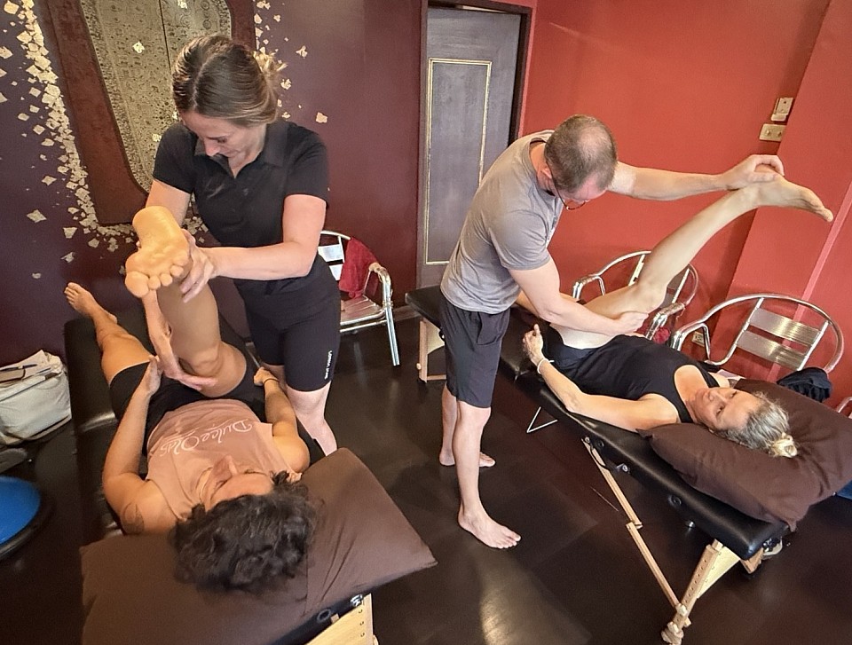
To practice sports-medicine–based manual therapy at a professional standard, a therapist must understand biomechanics, functional anatomy, joint mechanics, and the layered myofascial structure. This knowledge transforms structured assessment into a precise orthopedic-grade evaluation rather than a surface-level routine. While relaxation massage has value, the RSM approach requires interpreting how posture, fascia, joints, and neural structures interact—and using that analysis to deliver targeted, effective intervention.
Understanding the Human Body in a Clinical Context
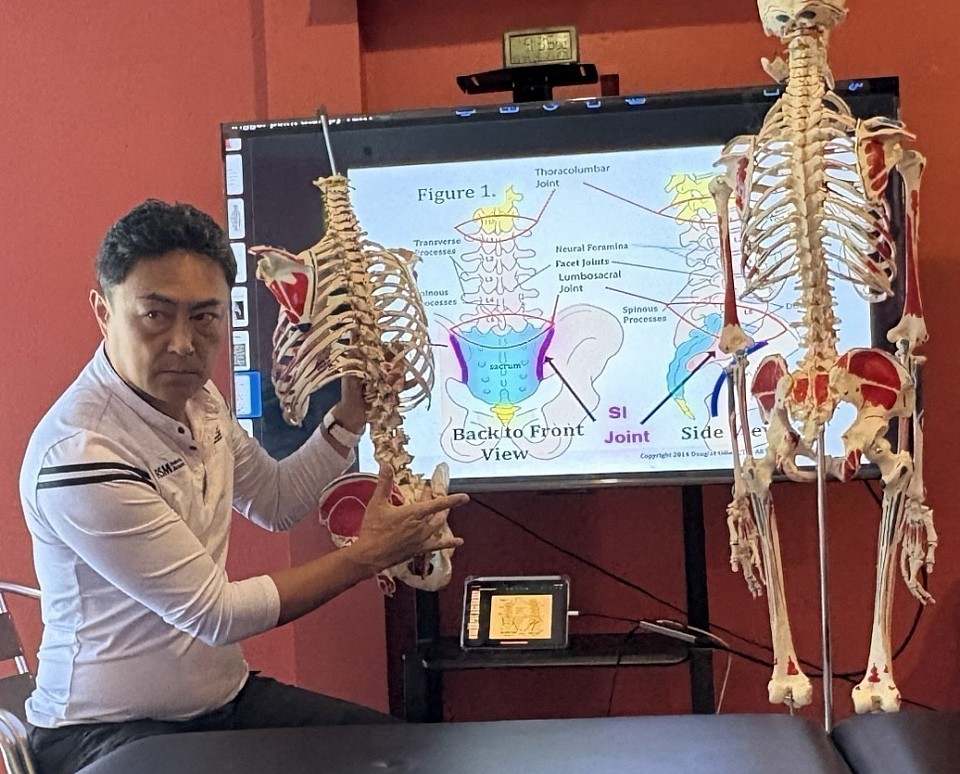
For a professional massage therapist, the body is never treated as a single structure but as an interconnected musculoskeletal system in which joints, fascia, muscles, and neural elements influence one another during movement. At RSM International Academy, this understanding is developed through 500–700 high-resolution clinical images used in each course to teach the three-dimensional layering of the body—skin, superficial fascia, deep fascia, skeletal muscle, tendons, ligaments, and bone. This detailed visual education enables practitioners to recognize how these layers behave under load and how dysfunction arises within the kinetic chain.
True clinical practice requires the ability to visualize structures beneath the skin and interpret how fascial tension, joint mechanics, and neural sensitivity interact. This is the core of advanced clinical palpation taught at RSM. When a client presents with low-back symptoms, the source is rarely a single “tight muscle.” Instead, the issue may stem from lumbar facet joint mechanics, pelvic alignment, deep posterior-chain fascial tension, or neural irritation such as Superior Cluneal Nerve–related low-back pain, which is frequently associated with Maigne’s Syndrome (Thoracolumbar Junction Syndrome)—a condition originating from dysfunction at the T12–L1 region that refers pain toward the iliac crest.
Grounding in physiology and structural function allows therapists to select interventions with precision rather than guesswork. By linking palpation findings to kinetic-chain mechanics and regional interactions, practitioners identify the true mechanical origin of dysfunction and deliver manual therapy consistent with modern sports-medicine standards. This integration of biomechanical reasoning and image-driven education is what makes RSM’s methodology unique within global manual-therapy training.
Mastering Anatomy for Better Client Outcomes
At RSM International Academy, the cornerstone of effective manual therapy is the ability to understand exactly what structure you are treating and how that structure behaves under load. Our sports-medicine curriculum goes beyond general charts and requires students to study origins and insertions at a clinical level—how each muscle attaches to bone, how force travels through these attachment sites, and why tension commonly builds at these anatomical entheses. This precision allows practitioners to locate true pain generators instead of treating only the superficial muscle belly.
Equally critical is the study of insertions and actions, which gives the therapist a clear understanding of the muscle’s line of pull. By analyzing how a muscle creates movement through its concentric, eccentric, and isometric phases, students learn how to release or stretch tissue in alignment with its mechanical vectors. For example, knowing the functional action pattern of the biceps femoris—or the exact insertion of the supraspinatus—allows the practitioner to design interventions that directly reduce impingement, restore joint centration, and reduce compensatory load across the kinetic chain.
This integration of origins & insertions with insertions & actions transforms a massage session into a strategic clinical process rooted in biomechanics, functional anatomy, and sports-specific demands. Rather than guessing, RSM-trained therapists make interventions with anatomical accuracy, predict how tissues will respond to pressure, and adjust techniques based on joint mechanics, fascial tension lines, and neural responsiveness. This is the level of precision that elevates treatment outcomes in pain reduction, posture correction, and performance optimization.
Applied Therapy Techniques and Functional Movement
Anatomy is not simply the study of muscles in isolation; it is the study of how those muscles coordinate movement, posture, and force transmission through the kinetic chain. At RSM International Academy, we integrate functional anatomy with manual therapy by teaching students to understand how structure and motion interact. Instead of memorizing charts, we use real clinical cases to analyze how pain is produced—what movement triggers symptoms, which tissues are overloaded, and how biomechanics shape the client’s presentation. This clinical context allows practitioners to link anatomy directly to real-world problems.
When students understand the kinetic chain, they see that neck pain may stem from thoracic dysfunction, scapular mechanics, joint-capsule restriction, myofascial tension, or neural irritation. This deeper perspective changes the pressure and direction of every stroke. In courses such as Deep Tissue Massage and Sports Medicine Massage, we emphasize that effective deep-tissue work is never about force. It requires sinking through anatomical layers with precision—guided by the structure, the tissue barrier, and the functional relationship between joints and fascia.
Using correct techniques protects both the client and the therapist. By aligning your body mechanics with the client’s anatomical planes, you avoid wasting time with ineffective pressure or injuring your own fingers and wrists. Instead of “pushing,” you learn to “sink” into tissue where anatomy indicates separation or restriction. Functional-anatomy-based manual therapy creates mutual benefit: the client receives accurate, rapid clinical results, and the therapist works efficiently with minimal strain. This philosophy underpins all training at RSM International Academy.
The Role of Massage Therapy in Pain Management
At RSM International Academy, our system is built from over 25 years of clinical experience—not from copying textbooks or spa-style routines. Manual therapy begins with understanding why the body loses balance: why muscles tighten, why the pelvis shifts, and why pain develops. We train practitioners to analyze functional anatomy, kinetic-chain behavior, joint mechanics, and myofascial tension before a single stroke is delivered. This approach transforms treatment into precise, targeted work based on real anatomical reasoning, not guesswork.
In our courses—Deep Tissue Massage, Sports Medicine Massage, and Neuro-Myofascial Release—therapists learn to identify whether dysfunction is muscular, fascial, joint-related, or neurological. They study origins and insertions, insertion actions, movement vectors, and how tissue behaves under load. Every technique, pressure angle, and stroke direction is chosen to restore joint mechanics, normalize movement patterns, and reduce pain efficiently. The result is a level of clinical accuracy that simply cannot be achieved through generic massage training.
Because of this depth, RSM attracts physiotherapists, Pilates instructors, medical doctors, and active clinical practitioners—who consistently make up 30–40% of each class. These professionals come not for relaxation techniques but for sports medicine–based manual therapy that directly upgrades their work in hospitals, clinics, and performance settings. Our Google Maps reviews reflect exactly why they value RSM: practical, evidence-grounded training that delivers immediate results in pain reduction, posture correction, and optimized movement.
Elevating the Standard of Care
At RSM International Academy, we teach therapists to understand how muscles, joints, fascia, and nerves are supposed to move—what proper mechanics look like, and how pain emerges when these systems fall out of sync. Whether it is muscular tension, joint-capsule dysfunction, fascial restriction, or neural mobility issues, we link every clinical problem to its biomechanical cause. Students learn through a sports-medicine framework, combining functional anatomy with precise hands-on manual therapy.
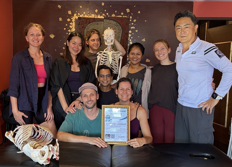
Our academy remains intentionally small—maximum seven students—because palpation accuracy and structural understanding determine the entire quality of manual therapy. Without understanding structure and function, “muscle release” becomes nothing more than a routine anyone can perform. But when a therapist understands the anatomical architecture, even a few seconds of contact can reveal tissue behavior, movement dysfunction, and the true mechanism of pain. This is what defines medical-grade manual therapy and separates top practitioners from technicians.
At RSM, we eliminate the outdated belief that “strong pressure equals effective treatment.” Instead, we train therapists to intervene with anatomical precision—guided by biomechanics, functional anatomy, and kinetic-chain reasoning. Understanding how muscles, joints, fascia, and nerves interact in real movement is what leads to meaningful change in pain, posture, and performance. This is the foundation of true clinical massage and the core philosophy of RSM.
- Hironori Ikeda, MSc Sports Medicine
Neurodynamics & Sports Biomechanics Specialist
Reference
1) Practicing Sports Massage. Massage Therapy Journal, May 2011. This article emphasizes that therapists working in sports settings must possess advanced skills in anatomy, pathology, orthopaedic assessment, and biomechanics.
2) The Ultimate Guide to Sports Massage: Techniques, Benefits, and Expert Tips. Massage Company Blog, 2023. This guide explores the deep link between sports massage, anatomy, physiology, and biomechanics — aligning strongly with your message about “functional anatomy → manual therapy.”
Sports massage course students at RSM international academy
ITBS and the Lower Cross Kinetic Chain: Beyond Lateral Knee Pain
Kinetic chain assessment and myofascial release
I frequently encounter cases of lateral knee pain labelled as Iliotibial Band Syndrome (ITBS), but in truth the problem often begins far from the knee. The chain typically starts with pelvic anterior tilt – a hallmark of a lower-cross syndrome. That anterior tilt increases lumbar lordosis, promotes femoral internal rotation, and lays the foundation for tension. In many clients I estimate that 60-70% of the tension on the iliotibial tract originates from the Tensor Fasciae Latae (TFL). From there the chain continues: TFL → fascial linkage across the lateral thigh → insertion around Gerdy’s tubercle → lateral knee load.
When the lateral thigh fascia – which covers from the iliac crest, runs outside the femur and wraps around the knee – loses its glide, the patella and surrounding structures suffer. In older adults especially the patellar fat pad may become fibrosed, compounding lateral knee pain. Skeletal alignment variations such as genu valgum (X-legs) or genu varum (bow-legs) also shift load onto the lateral chain. For athletes, foot pronation, femoral anteversion/retroversion and soft running shoes can aggravate the chain.
At RSM International Academy our Deep Tissue Massage and Remedial Massage courses address these mechanisms. Students learn to evaluate pelvic tilt, femoral torsion and pronation/supination mechanics — not just treat the knee. Because ITBS is better understood as a chain issue, not just a local band problem.
- Hironori Ikeda, MSc Sports Medicine
Neurodynamics & Sports Biomechanics Specialist
Reference :
1) Falvey EC, Clark RA, Franklyn-Miller A et al. “Iliotibial band syndrome: an examination of the evidence behind a number of treatment options.” Scand J Med Sci Sports. 2010;20(4):580–587.
2) Bonoan M. “Iliotibial band syndrome: Current Evidence.” Int J Sports Phys Ther. 2024.
