RSM Blog: Sports Medicine and Massage Insights
Managing Fatigue After Massage Work: A Sports Medicine Guide
Sports medicine massage course
Fatigue experienced after a thoroughly performed deep tissue massage extends beyond typical exertion-related tiredness or the lethargy associated with illness. Clients often describe this sensation as feeling "boneless," "wrung out," or as if their body has finally ceased internal conflict. Practitioners specializing in sports medicine are well-acquainted with this response and recognize how inadequately it is often explained. Clients frequently receive vague reassurances about "toxins being released" or are simply advised to hydrate—explanations that neither satisfy physiotherapists nor athletic trainers seeking a deeper understanding of the physiological processes involved.
The Autonomic Shift: Understanding Post-Massage Fatigue
The primary cause of fatigue following massage is neurological rather than muscular.
Moderate-pressure massage consistently triggers a parasympathetic nervous system response, which can be quantified through heart rate variability analysis. Within minutes of treatment, there is a shift toward increased vagal efferent activity and a reduction in sympathetic dominance. The body transitions from an active operational state to its rest-and-digest mode: cortisol and norepinephrine levels decrease, while serotonin and dopamine increase. For individuals experiencing chronic stress or undertraining, this shift represents a significant physiological downregulation rather than a subtle adjustment. The body, often for the first time in hours or days, genuinely ceases its defensive tension.
This state is subjectively perceived as exhaustion but is not pathological; rather, it signifies a healing phase, a distinction of clinical importance. Practitioners who misinterpret post-massage fatigue as an adverse effect may reduce pressure or shorten sessions to avoid inducing it. In sports medicine, however, this is typically counterproductive. The autonomic shift is precisely the condition that facilitates effective tissue repair and recovery. Educating clients that post-session tiredness often indicates therapeutic success, rather than a complication, constitutes a valuable clinical intervention.
Metabolic By-Products, Lactic Acid, and Debunking the Toxin Myth
The pervasive narrative of "toxin release" in massage culture persists because it provides clients with an intuitive explanation for a genuine physiological phenomenon. In reality, massage modifies circulation and lymphatic flow, facilitating the movement of normal metabolic by-products through tissues. These substances include lactate, hydrogen ions, and inflammatory mediators such as cytokines and prostaglandins. None qualify as toxins in any clinical context; rather, they are routinely produced and cleared by the body.
Lactic acid’s role is frequently misunderstood. Lactate functions as both a fuel substrate and a signaling molecule, not as a waste product requiring elimination. Following intensive massage on heavily loaded muscle groups, transient elevations in local metabolite concentrations may occur as compressed tissues are reperfused. This, combined with localized micro-inflammatory responses, can produce flu-like symptoms reported by some clients after deep sessions, including mild achiness, limb heaviness, or slight nausea. These symptoms are self-limiting, typically resolving within 12 to 24 hours, and are most common after initial sessions or prolonged intervals between treatments.
Hydration remains a sound clinical recommendation post-massage, independent of the toxin narrative. Adequate fluid intake supports renal clearance of metabolic by-products, maintains optimal blood viscosity during increased circulation, and ensures efficient lymphatic function. The physiological rationale for this advice is robust; only the terminology requires refinement.
Deep Tissue Massage, Muscle Soreness, and the Healing Process
Deep tissue massage warrants a distinct recovery framework. When structural manipulation disrupts longstanding adhesions or restores perfusion to ischemic tissues, the body initiates an inflammatory cascade. Creatine kinase levels may transiently rise in the bloodstream, paralleling responses observed after eccentric exercise. This explains why muscle soreness following deep tissue massage often peaks 24 to 48 hours post-treatment, mirroring the trajectory of delayed onset muscle soreness. Such discomfort signals physiological adaptation rather than injury.
In sports medicine, this understanding informs session scheduling. Aggressive deep tissue work within 48 hours of competition may induce soreness and temporary reductions in peak force output. Consequently, clinical consensus favors lighter, circulation-enhancing massage during the 48 to 72 hours preceding events, reserving deeper structural interventions for post-event recovery or training periods well separated from competition. The Deep Tissue Massage Course at RSM provides comprehensive guidance on these considerations, including client physiological assessment prior to sessions and integration of deep tissue techniques within periodized training plans.
When Persistent Fatigue Post-Massage Warrants Further Evaluation
Typically, post-massage fatigue resolves within 24 hours. Fatigue extending beyond this timeframe signals a different clinical concern.
Clients who are immunocompromised, significantly deconditioned, or experiencing overreaching may exhibit disproportionate recovery demands following standard sessions. Prolonged exhaustion in these cases reflects an exceeded adaptive capacity rather than an inherent issue with massage therapy. Appropriate management involves recalibration—shorter sessions, reduced intensity, or adjusted timing—rather than discontinuation of treatment.
Clinicians should monitor for patterns such as fatigue persisting beyond 24 hours accompanied by disrupted sleep or ongoing muscle soreness; clients who report worsening symptoms on days two or three post-treatment instead of improvement; and emotional disturbances such as irritability or heaviness extending beyond the treatment day, often indicative of elevated pre-existing stress interacting with autonomic nervous system responses to intensive therapy.
Effective Strategies for Managing Post-Massage Fatigue and Recovery
Conventional post-massage care advice tends to be generic. A more nuanced approach recognizes the distinct mechanisms involved.
Recovery of the nervous system is paramount in the hours following treatment. Clients experiencing parasympathetic dominance benefit from gentle movement rather than immediate engagement in cognitively demanding tasks or stimulant consumption. Light ambulation promotes venous return and lymphatic drainage without reactivating the sympathetic nervous system.
Muscle recovery after deep structural work aligns with exercise recovery principles. Adequate protein intake supports repair of mechanically stressed tissues. Quality sleep during the night following intensive sessions is critical, as this period consolidates soft tissue remodeling and resolution of inflammation. Cold-water contrast therapy may alleviate localized soreness and expedite inflammatory recovery.
Hydration underpins all these processes, and for athletes, maintaining electrolyte balance is essential following sessions involving significant flushing of heavily trained muscle groups.
The Importance of Precise Communication in Recovery
The terminology employed by therapists profoundly influences clients’ perceptions of their recovery. Framing the post-massage autonomic shift in neurological terms rather than invoking toxins provides clients with a clear, anxiety-reducing framework that enhances adherence to post-care recommendations. Physiotherapists appreciate this precision; athletic trainers relate it to session planning; yoga instructors recognize the parasympathetic state akin to savasana.
At RSM, we dedicate significant attention to this aspect. Clinical communication is a critical skill, not a peripheral one. In sports medicine, the ability to accurately articulate post-treatment physiological processes determines how massage is perceived by colleagues and whether it becomes an integral component of recovery protocols or remains marginal. Practitioners who anticipate client experiences within 24 to 72 hours post-treatment, communicate these effectively, and adapt their approach accordingly, establish enduring clinical credibility. This level of precision is our professional objective.
Stretching After Trigger Point Release: Why the Sequence Is Everything
Trigger point massage course
I often see therapists working with trigger points focus their attention on the compression itself: the depth of the contact, the pressure vector, the response of the taut band, the referred pain pattern confirming location. That attention is warranted. But the compression is only half the treatment. What the muscle needs in the sixty seconds immediately after a trigger point releases is what most protocols underspecify, what most sessions rush past, and what the physiology actually demands. Stretching after the release of a trigger point is not a cool-down gesture. It is the mechanism by which a transient neurological event becomes a durable mechanical change.
What Actually Happens at the Trigger Site
The current working model describes the myofascial trigger point as a cluster of hypercontracted sarcomeres concentrated near a dysfunctional motor endplate. When local ATP is depleted through sustained low-level contraction, eccentric overload, or acute trauma, calcium accumulates in the cytosol and sarcomeres lock in a shortened state. The contracted zone compresses local capillaries, restricting blood flow and creating a localized ischemic environment. The tissue becomes acidic, with pH values in active trigger points recorded as low as 4.5 in microdialysis studies by Shah and colleagues. That chemical milieu, rich in substance P, bradykinin, and inflammatory cytokines, sensitizes local nociceptors and sustains the pain cycle.
The structural consequence is a palpable contraction knot: a segment of muscle fiber with maximally shortened sarcomeres, flanked by the abnormally stretched portions of the same fiber pulled taut to accommodate the shortening. The taut band running through the muscle is not uniformly contracted. It is a system under internal mechanical tension, with some sarcomeres in crisis and others in compensatory overstretch. Releasing a trigger point through sustained compression or dry needling disrupts that biochemical environment and allows the locked sarcomeres to let go, but it does not automatically restore the fiber to its resting length. The surrounding connective tissue imposes structural inertia. Stretching is what addresses that inertia.
The Plasticity Window: Where Pain Relief Becomes Structural Change
Travell considered post-release stretching essential, and her spray-and-stretch technique was built on the principle that neurological gating and tissue lengthening must be combined for durable deactivation. The same logic applies to manual release: compression desensitizes the point and permits a brief window during which muscle tissue accepts lengthening it would normally resist.
Immediately after release, local blood flow increases in a reactive hyperemia that washes metabolic waste from the area and restores pH toward neutral. Nociceptor firing diminishes. Protective muscle guarding drops. The fascial tissue, having been subjected to sustained mechanical load during compression, exhibits a transient reduction in stiffness. These effects are time-limited, probably in the range of one to three minutes. A study examining ischemic compression followed by PNF stretching in subjects with latent trigger points in the pectoralis minor found the combination produced significantly greater increases in muscle length than compression alone. The window is real, and it closes.
Which Stretches Work Best After Release, and Why
Three approaches have the most clinical support after trigger point therapy: passive sustained stretch, post-isometric relaxation (PIR), and contract-relax PNF.
Passive sustained stretch is appropriate when tissue is acutely sensitized or when the released muscle sits in a region like the neck or lower back where aggressive loading carries risk. The muscle is taken slowly to the first point of resistance and held, allowing the viscoelastic properties of the connective tissue to respond. PIR takes a different route: the client performs a brief isometric contraction of the just-released muscle against the therapist's resistance, then relaxes into a deeper passive stretch. The contraction activates the Golgi tendon organs, inhibiting the muscle's own motor neurons and creating a window of enhanced receptivity to lengthening. Contract-relax PNF operates on similar neurological principles with greater targeting precision, and research comparing these methods after ischemic compression found it produced the greatest improvements in muscle length. For most clinical scenarios, PIR immediately post-release, followed by the client actively moving through full range of motion three times, captures both the neurological and mechanical benefit.
Neck, Back, and Shoulder: Applying Trigger Point Releases Where It Matters Most
The muscles that present most frequently with active trigger points, including the upper trapezius, levator scapulae, quadratus lumborum, and infraspinatus, each carry specific post-release stretching considerations.
In the neck and upper shoulder, the most common error is stretching before the release is complete. Sustained pressure on an upper trapezius trigger point produces referred pain to the lateral neck and temporal area; the release is signaled by a softening under contact and a reduction in that referral. Stretching before that signal appears meets a muscle still in guarded state. In the lower back, the quadratus lumborum's trigger points refer pain to the iliac crest and buttock, and because the QL is temporarily neurologically inhibited immediately after release, post-release stretching should be done in a side-lying position where gravity assists rather than a standing stretch that demands spinal stability from the very muscle being targeted.
Self-Massage, Trigger Point Tools, and Home Practice
The benefit of trigger point work extends beyond the treatment table only when clients understand the sequence. Self-massage trigger point tools allow clients to approximate ischemic compression at home, but most cannot simultaneously monitor pressure intensity, sustain it appropriately, and then transition into the subsequent stretch without explicit training. Clients who have been shown the specific stretch for each treated muscle, and who have practiced the transition in session, replicate it at home with genuine clinical usefulness.
A practical home sequence: sustained pressure on the target muscle until local and referred pain begins to diminish, typically within 60 to 90 seconds, followed within 30 seconds by a passive stretch held for 30 to 60 seconds, then active movement through full range three to five times. Frequency of two to three times daily for acute presentations, reducing to once daily as symptoms stabilize. The order is not negotiable. Stretching before the compression achieves less: the guarded tissue resists, and the client experiences discomfort without proportionate pain relief.
Pressure, Duration, and the Completeness of a Point Release
The effectiveness of any subsequent stretch depends on what was actually achieved during compression. A superficial or brief point release that does not sustain long enough for referred pain to diminish leaves the muscle in a partially guarded state, and the plasticity window that follows is narrower as a result. A complete release is signaled by a palpable softening of the taut band, a reduction in the referred pain pattern, and visible relaxation of the muscle. These changes are not instantaneous: depending on chronicity and depth, a release may require anywhere from 30 seconds to several minutes.
I see this consistently with therapists who are technically accurate but clinically impatient. The contact position is correct. The vector is right. But the hold ends before the tissue has responded, which means the subsequent stretch meets a muscle that is only partially ready to accept it. The hold ends when the tissue signals readiness, not when the clock reaches a preset number.
Building Muscle Releases Into a Complete Session Protocol
Integrating stretching after trigger point releases requires sequencing awareness more than extra time. A release followed immediately by a stretch takes perhaps ninety seconds longer per site. Across a session addressing four or five active trigger points, that is seven or eight additional minutes. The return, in durable range of motion, reduced pain recurrence, and reinforced treatment value, justifies it.
At RSM, this is the principle we return to in the Trigger Point Therapy Course: technique is the entire sequence, from palpation to release to stretch to active movement, not just the compression. The compression deactivates the trigger point. The stretch consolidates the change. Skip either, and the result is diminished.
Current Research in Remedial Massage: What the Evidence Really Means for Clinical Practice
Remedial massage Course
There is a particular kind of frustration that comes from watching a field you respect be misrepresented, either by its critics or its most enthusiastic advocates. For those of us working at the intersection of sports medicine and massage, that frustration has become familiar. Massage is one of the oldest physical interventions in human medicine, and over the past two decades it has accumulated an evidence base substantial enough to deserve careful reading. What the most recent large-scale reviews have done is force a more honest and ultimately more useful conversation about what massage therapy research can and cannot tell us.
A Large Evidence Base With a Significant Catch
In July 2024, JAMA Network Open published the most comprehensive evidence map of massage therapy for pain produced in recent years. Researchers at the Veterans Health Administration systematically reviewed 129 systematic reviews covering massage for painful adult health conditions across 13 categories, from chronic low back pain and neck pain to cancer-related pain and fibromyalgia. Of those reviews, only 17 met criteria for the evidence map. The headline finding was blunt: not a single conclusion was rated as high-certainty evidence. Seven conclusions reached moderate certainty, and all seven supported the position that massage therapy is beneficial for pain.
This is not a finding that massage therapy does not work, and that distinction matters enormously. Low certainty means our confidence in the effect estimate is limited, not that the effect is absent. The problem is methodological. When research pools Swedish relaxation massage, myofascial release, deep tissue work, and specific clinical massage protocols into a single intervention category, the results become difficult to interpret for anyone trying to make evidence-based decisions in practice. The field of remedial and therapeutic massage operates with considerably more clinical nuance than most research designs currently capture.
What Moderate-Certainty Massage Therapy Research Actually Shows
The seven moderate-certainty conclusions covered massage for labor pain, chronic low back pain, neck pain, and several other musculoskeletal conditions. In each case, massage had a beneficial association with reduced pain intensity compared to control conditions. For chronic pain populations, that is clinically significant: the effect was consistent enough across independent studies and participant groups to survive rigorous quality assessment.
The pattern tells a coherent story even if it does not yet tell a complete one. Massage therapy produces meaningful clinical benefits for pain, stress, and physical function across a wide range of conditions. The evidence base is not too weak to act on; it is too inconsistently structured to generate the high-confidence conclusions that systematic reviews are designed to produce. A 2025 meta-analysis in the Journal of Clinical Nursing, covering 36 randomized controlled trials with 3,671 participants, found that massage therapy significantly improved pain, quality of life, and anxiety in cancer care populations, with a pooled pain-reduction effect size of -0.51, yet evidence certainty remained very low for the same structural reasons.
The Neuroscience of Therapeutic Massage and What It Explains
While the clinical trial literature navigates its methodological constraints, the neuroscience of therapeutic massage has been advancing with considerably more traction. This is where current research is producing its most practically relevant findings.
Oxytocin, Pressure Receptors, and the Spinal Cord Circuit
A 2026 preprint combining human and mouse models provided what may be the most mechanistically complete explanation yet of how massage modulates pain and touch reward simultaneously. The research demonstrated that massage triggers oxytocin release, which acts at the spinal dorsal horn through a state-dependent circuit, modulating ascending pain pathways by acting on both excitatory and inhibitory spinal neurons. In human participants, well-being responses to massage correlated directly with endogenous oxytocin levels. This explains why moderate-pressure massage produces qualitatively different physiological effects than light touch. Earlier work by Tiffany Field and colleagues at the Touch Research Institute established that moderate pressure increases vagal activity, reduces cortisol, and elevates serotonin and dopamine, while light touch tends to produce sympathetic arousal rather than parasympathetic activation. The spinal oxytocin circuit data provides the upstream mechanism for what had previously been observed only at the level of autonomic and endocrine outcomes.
Neuroimaging has added further structural detail. Resting-state fMRI in chronic pain populations has documented normalization of connectivity within default-mode and salience networks following multi-session massage interventions, with changes that persist at delayed follow-up. The implication is that therapeutic massage produces neuroplastic changes, not merely transient physiological responses.
Clinical Massage in Oncology: What the Research Reveals
Oncology is one of the most important clinical contexts for massage therapy. Cancer and its treatments produce pain, anxiety, fatigue, and damage to musculoskeletal health through mechanisms that conventional pharmacological care often cannot fully address. The 2025 Journal of Clinical Nursing meta-analysis, alongside a 2024 analysis focused on post-surgical breast cancer patients, confirmed significant reductions in pain and anxiety following massage therapy. Yet both acknowledged high risk of bias in most included studies. The gap between clinical signal and rated evidence certainty is a direct consequence of trial design: heterogeneous participant populations, under-specified massage protocols, and inconsistent outcome measurement. Improving that requires massage therapists to be more involved in research planning and requires researchers to develop cleaner taxonomies of massage interventions that actually reflect clinical practice.
What This Means for Health and Clinical Practice
From where I stand, having trained therapists in the evidence-based application of remedial and clinical massage for many years, the recent literature communicates something specific and actionable even when it does not deliver the definitive proof that headlines prefer.
The physiological case for massage therapy as a legitimate health intervention is now substantially better supported at the mechanistic level than it was a decade ago. We understand how pressure magnitude determines autonomic response. We have the oxytocinergic spinal circuit. We have neuroimaging data showing that multi-session massage effects extend into resting-state brain connectivity. These mechanistic findings give clinical practice a defensible foundation even where large trial evidence remains limited.
The most important practical insight from the recent literature is the dose-response relationship. Moderate pressure for appropriate durations produces parasympathetic activation, oxytocin release, and cortisol reduction. Light pressure does not produce the same physiological profile. Session frequency matters too: the neuroplastic and sustained autonomic changes appear to require multiple sessions, not single assessments. Therapists working with clients presenting chronic pain, musculoskeletal health complaints, or oncology-related symptoms have more evidentiary support than the headline certainty ratings suggest. Calibrating that evidence conversation accurately, without overstating certainty, is itself a clinical skill.
The therapist who understands the oxytocinergic pathway, who knows what the JAMA Network Open evidence map showed and what it did not show, is a better clinician and a better advocate for the field. That combination of technical depth and research literacy is what our Remedial Massage Course is designed to develop. The research is not yet where it needs to be. The clinical case, understood carefully, is considerably stronger than the headlines suggest.
Differences Between Active and Passive Myofascial Release: Choosing the Right Tool
Dynamic Myofascial release Course
Therapists often encounter the active/passive distinction early in their training and file it away as a simple binary: active means the patient moves; passive means they lie still. That framing is technically accurate and almost entirely useless. The real clinical question isn't what the patient does with their body; it's what physiological mechanism you are trying to engage, and whether your chosen release therapy approach matches the tissue state in front of you. Getting that match wrong doesn't just produce slower results. In certain cases, it produces no results at all, and the patient's pain remains exactly where it started.
Why Passive Release Techniques Address Chronic Myofascial Pain Differently
In passive myofascial release, the patient does nothing. The tissue is taken to a point of restriction and held for 90 seconds to three minutes, waiting for fascial creep rather than a muscle guarding reflex. The absence of patient effort creates a neurological environment in which competing proprioceptive signals are quieted, allowing the therapist to detect subtle restriction vectors that active contraction would otherwise mask.
This approach suits chronic, global restrictions where densified and adherent myofascial tissues require sustained mechanical loading to undergo thixotropic change. A soft tissue that has been restricted for years will not respond to a 30-second pass of moderate pressure. There is also a psychophysiological benefit: a patient asked to do nothing but breathe is more likely to downregulate sympathetic tone, which meaningfully changes the tissue's response to the same level of applied pressure. For patients managing myofascial pain with significant central sensitization, the passive environment is not a compromise but a genuine therapeutic variable, and the therapy itself becomes part of the neurological reset.
Active Release and the Neuromuscular Logic Behind It
Active release operates on different logic. The tissue is taken to a shortened position, the therapist establishes specific contact, and the patient moves through a lengthening arc under load. The shear force generated between the therapist's contact point and the moving tissue disrupts adhesions between layers, not by waiting for a creep response, but by creating controlled friction as the muscle elongates. To release myofascial adhesions through movement rather than sustained hold is the defining distinction of this approach.
Active Release Technique, widely used across chiropractic, sports medicine, and physiotherapy, formalizes this into a diagnostic and treatment algorithm. Practitioners identify zones of restriction by evaluating texture, tightness, and movement, then apply contact-and-move protocols targeting the cumulative trauma cycle: the progressive accumulation of micro-injuries that reduces tissue quality without producing any single obvious injury event. For athletes, this approach offers something passive work cannot replicate as efficiently. Eccentric lengthening under the therapist's hands trains the tissue in the same mechanical conditions it will face in training, building the functional tolerance that passive release alone does not produce.
Reading Tissue Readiness: When Each Active or Passive Approach Fits
I have found, working with athletes and physically active patients over many years, that the most common clinical error is not choosing the wrong technique; it is applying the correct technique at the wrong time. Passive release applied during an acute inflammatory response will irritate the tissue further. Active release applied before a healing soft tissue injury has sufficient tensile strength may disrupt the early collagen matrix. Understanding the passive active continuum is therefore not a theoretical exercise; it is a practical release technique decision made at the table, informed by what the tissue is telling you.
A more useful framework than "active versus passive" is tissue readiness. In the acute phase, passive approaches protect the structure while addressing the fascial restrictions already forming around the injury site. As healing progresses, active release begins to address the specific adhesions forming between recovering tissue layers, the ones that, left untreated, become the chronic limitation that reduces performance months later.
Key factors in that clinical decision include the acuity of the injury, the patient's pain pressure threshold at the target site, whether the primary limiting factor is tissue densification or scar tissue adhesion, and whether the patient can produce controlled movement through range without compensating. Pain that sharpens with active loading is often the clearest signal to stay passive longer.
How RSM Teaches the Release Technique Spectrum
Within the RSM Myofascial Release Course, we teach both approaches as complementary tools within a coherent treatment architecture. A session often opens with passive work: guarding reduces, global tone settles, and the therapist's proprioceptive picture of the fascial system clarifies. Active release can then be introduced with greater precision because the surrounding soft tissue has already been addressed.
What changes between approaches is not just mechanics; it is what you are asking of the patient's nervous system. Passive work says: let go, I will find the restriction. Active work says: move, and let the movement reveal what needs to change. Both are asking for a release; they are simply using different physiological levers. Clinical sophistication lies in knowing which lever belongs to which moment, and in having the palpation skill to feel when that moment has arrived.
The Science Behind Trigger Point Formation: From Energy Crisis to Central Sensitization
Trigger point therapy course
Finding trigger points, compressing them, and releasing them is well within the reach of most experienced therapists. Far fewer can explain, with precision, what they actually are at a cellular level. That gap matters. Not because clinical intuition is inferior to molecular biology, but because understanding the mechanism behind the pathology changes how you think about treatment, which patients you prioritize for which techniques, and how confidently you discuss myofascial pain with the sports physicians and physiotherapists on your team.
What Travell and Simons Established
Janet Travell and David Simons spent decades mapping myofascial pain patterns and codifying what they called myofascial trigger points. Their foundational insight was that a trigger point is not simply a tender spot. It is a hyperirritable nodule within a taut band of skeletal muscle with a predictable referred pain pattern, reproducible on compression, and capable of generating both local and distant symptoms.
Simons and Travell placed trigger points on a spectrum. An active trigger point produces spontaneous pain that typically refers to a distant site. A latent trigger point is only painful when mechanically stimulated, but it still restricts range of motion and alters motor recruitment. The distinction matters clinically: latent points are far more prevalent than active ones, they can convert to active status under physical or psychological stress, and they represent genuine neuromuscular dysfunction even in the complete absence of pain.
Their first mechanistic explanation, the energy crisis hypothesis, proposed that an overloaded motor endplate releases excessive acetylcholine (ACh), producing abnormal sustained depolarization and persistent sarcomere contraction around the endplate zone. Without adequate ATP to return calcium to the sarcoplasmic reticulum, the muscle cannot relax. Metabolic demand increases, capillary compression reduces blood flow, and the local environment becomes hypoxic and ischemic. The muscle cannot generate enough energy to break the contracture, so the contracture sustains itself.
This model explained the palpable taut band, the local tenderness, and why stretching or sustained compression might interrupt the cycle by mechanically elongating contracted sarcomeres and lowering ATP demand.
The Feedback Loop That Keeps the Trigger Active
The integrated trigger point hypothesis, expanded in 2004 by Gerwin, Dommerholt, and Shah, revealed a more complex cascade. Ischemia does not simply deplete energy; it creates an acidic local environment. That acidity inhibits acetylcholinesterase (AChE), the enzyme that ordinarily clears ACh from the synaptic cleft. With AChE suppressed, ACh lingers and endplate activity remains abnormally elevated. Acidic conditions also stimulate the release of calcitonin gene-related peptide (CGRP), which further inhibits AChE and upregulates ACh receptors on the postjunctional membrane, amplifying the signal further still.
The feedback loop closes tightly: ischemia drives acidosis, acidosis amplifies ACh signaling, amplified ACh signaling intensifies sarcomere contraction, and intensified contraction worsens ischemia. This is what separates trigger point formation from ordinary post-exercise soreness. In most muscle, protective mechanisms interrupt this cascade before it becomes self-sustaining. In trigger point formation, they fail.
Sympathetic nervous system (SNS) activity adds a significant layer. Adrenergic receptors at the neuromuscular junction and in skeletal muscle respond to sympathetic activation by constricting capillaries and potentiating abnormal endplate firing. Patients under sustained psychological stress, or with chronically elevated autonomic arousal, appear meaningfully more susceptible to trigger point formation independent of biomechanical loading. SNS activity is a perpetuating factor, not a secondary effect.
The Chemistry of Pain at the Point
The expanded hypothesis remained largely theoretical until Jay Shah and colleagues at the NIH developed a microdialysis technique capable of sampling interstitial fluid from living human muscle in real time. Inserting a fine needle into the upper trapezius of subjects with active trigger points, latent trigger points, and no trigger points, they produced the first direct biochemical portrait of myofascial pain at the tissue level.
Active trigger points showed significantly elevated concentrations of substance P, CGRP, bradykinin, serotonin, norepinephrine, TNF-α, IL-1β, and IL-6, alongside a measurably lower local pH. These are not incidental findings. Substance P and CGRP lower the activation threshold of muscle nociceptors, so stimuli that would normally be sub-threshold begin generating pain signals. Bradykinin directly activates nociceptors at very low concentrations. The cytokines contribute to peripheral sensitization and may drive structural tissue changes within the taut band over time.
Latent points showed an intermediate picture: sensitizing substances elevated above normal, but not to the degree found in active sites. This confirms that latent trigger points are not biochemically inert. They exist in a sensitized state that represents real tissue pathology, not merely a palpatory finding.
Shah's team also documented what followed a local twitch response (LTR), the brief involuntary contraction of taut band fibers that needling and some manual techniques reliably elicit. After an LTR, concentrations of substance P and CGRP dropped significantly at the active site, shifting the biochemical milieu toward the latent profile. This is one of the clearest mechanistic explanations for why techniques that reliably provoke an LTR tend to produce superior outcomes: they alter the chemistry that is driving peripheral sensitization, not just the mechanics of the tissue.
How Trigger Points Drive Central Sensitization and Referred Pain
The peripheral sensitization at an active point does not remain local. Sustained nociceptive input reaching the spinal cord reorganizes central pain processing. Dorsal horn neurons receiving prolonged input from the affected muscle segment become hyperexcitable: their activation thresholds fall, their receptive fields expand, and neurons that previously responded only to the directly affected segment begin responding to stimulation from adjacent regions.
This is the neurophysiological basis of referred pain. The characteristic referred pain patterns that Travell and Simons mapped so systematically are not a mystery; they are the predictable consequence of central sensitization in the dorsal horn. A trigger point in the infraspinatus generates anterior shoulder pain that can mimic rotator cuff pathology. A point in the deep gluteal muscles can reproduce sciatic symptoms. The therapist who understands this mechanism is far less likely to treat the site of reported pain and far more likely to locate the actual source.
CGRP plays a particularly important role in the shift from acute to chronic myofascial pain. It potentiates ACh activity at the motor endplate (perpetuating the peripheral trigger point) while also enhancing dorsal horn responses to substance P via the NK-1 receptor. This dual action at both peripheral and central levels makes CGRP a central mediator of why some patients develop widespread pain from what began as a localized muscle problem.
What This Means at the Table
I teach students at RSM International Academy to treat every trigger point as evidence about the state of the patient's entire system. The active point tells you something about the muscle's loading history, the patient's autonomic state, their recovery quality, and the degree to which central sensitization has already taken hold. A patient whose central sensitization is well established requires a fundamentally different approach than a healthy athlete recovering from training overload. Eliminating the peripheral source may be necessary but will rarely be sufficient on its own.
Myofascial pain syndrome driven by active trigger points is among the most common and underdiagnosed presentations in sports medicine. Patients with MPS present with diffuse, migrating pain that worsens under stress, does not resolve with rest as ligamentous injury would, and produces unremarkable imaging. The science behind trigger point formation gives the clinician a precise explanatory framework for these presentations.
In athletic populations, latent trigger points deserve particular attention. They suppress force output, alter recruitment patterns, and load adjacent structures in ways that produce recurring secondary injuries, without generating spontaneous pain. Identifying and treating latent points in these patients is genuinely prophylactic work.
The mechanistic picture also clarifies why manual techniques work when they work. Compression, muscle energy, and assisted stretching all aim to mechanically elongate contracted sarcomeres and restore capillary perfusion, operating on the same basic principle as dry needling. Knowing that the local environment at an active point is a genuine inflammatory milieu, and not merely a site of mechanical dysfunction, should sharpen how therapists think about technique selection, dosage, and treatment frequency.
Treating Trigger Points with Precision
RSM International Academy's Trigger Point Therapy Course is built on this mechanistic foundation. The curriculum covers the full evidence base for trigger point formation, assessment protocols for distinguishing active from latent points, the clinical significance of referred pain patterns across the major muscle groups, and hands-on technique grounded directly in the physiology described here.
Simons and Travell built the framework. The microdialysis research gave it biochemical substance. What we do at RSM is translate that into precise, reproducible clinical skill. The science behind trigger point formation is not background reading. It is the reasoning that separates a therapist who achieves durable results from one who applies pressure and hopes for the best.
Study Tips for Massage Students at the Advanced Level
Remedial massage course
Most of the massage study tips circulating online are aimed at entry-level learners trying to survive anatomy exams. Search any massage school forum and the advice is generic by necessity: take good notes, use flashcards, get enough sleep. For a physiotherapist, an athletic trainer, a doctor, or an experienced massage practitioner enrolling in an advanced clinical program, that advice is not wrong so much as it is beside the point. The challenge at this level is not absorbing new information; it is integrating a new clinical framework into an existing body of knowledge, habits, and muscle memory that are already well-established. That is a fundamentally different cognitive task, and it requires a different approach to study.
Why Experience Can Complicate Massage Therapy Learning
Experienced practitioners bring genuine advantages into any advanced program: clinical intuition, an understanding of tissue quality, and a feel for the body that no amount of classroom time can replicate. What experience can also bring, however, is entrenched habit. When you have applied a technique successfully for years, the nervous system has consolidated that pattern deeply. Introducing a new model, whether it is myofascial loading or a different approach to joint mechanics, means working against existing motor and conceptual schema.
Research in sports medicine consistently shows that skilled athletes learning a corrected movement pattern initially perform worse than beginners learning it fresh. The existing pattern competes. The same applies to advanced therapy students: your prior framework will resist the new one before it eventually integrates. The most effective response is to treat new material as a parallel system rather than a replacement. Study it on its own terms first, build the new structure separately, then look for the bridges.
The Dual Track: Separating Cognitive from Hands-On Therapy Work
Advanced massage therapy programs move between two fundamentally different kinds of learning: declarative knowledge (biomechanics, anatomy, pathology, assessment logic) and procedural knowledge (technique execution, palpation, clinical reasoning under real conditions). Conflating these in your study approach is one of the most consistent mistakes seen in students arriving with strong prior backgrounds.
Declarative content responds well to spaced repetition and active retrieval. Use index cards, brief written summaries, or practice tests you write for yourself. Force your brain to retrieve rather than re-read; re-reading feels productive but builds little lasting retention. Procedural knowledge, by contrast, builds through repetition on tissue. No study resources, however good, replace contact hours. For hands-on components of any massage course, short frequent sessions outperform long blocks: twenty minutes of focused technique work repeated daily will outperform a three-hour session on the weekend. If a class introduces a new assessment protocol, the goal is not to master it in the room but to rehearse it enough times across the following week that the movement becomes retrievable under real conditions.
Reading the Body Before Reading the Textbook
One of the more counterintuitive study strategies for advanced therapy students is to use the treatment table as a primary learning resource. Before reading the chapter on the rotator cuff, palpate the structures. Feel how tissue changes when the shoulder is internally versus externally rotated. Then read the chapter.
This sequence leverages the way the brain consolidates spatial and tactile information. When you subsequently encounter the anatomy in written or diagrammatic form, you are attaching a label to a sensation already encoded. The retention is substantially stronger. This is also how clinical massage reasoning actually functions: tissue information arrives first, and the conceptual framework organizes what the hands have found. The assessment sequences in our Remedial Massage Course are built on exactly this principle, with technique and tissue exposure preceding explanatory framework.
Study Tips for Massage: Prioritizing as a Working Clinician
Most students in an advanced program are also working clinicians. A realistic study plan is not built around maximizing hours; it is built around protecting the quality of a smaller number of hours. Prioritize material that bridges theory and clinical decision-making. In a remedial massage program, understanding the biomechanical rationale behind assessment tests matters more for long-term competence than memorizing test names. The names you can review before an exam; the reasoning, once genuinely understood, restructures how you assess every client for the rest of your career.
I started RSM because I believed there was a gap between what standard massage education offers and what clinical work at a sports medicine level actually demands. Every student who comes through this school brings prior knowledge that is genuinely valuable, and every one of them encounters material that challenges what they thought they already knew. The most reliable study tip I can offer any experienced practitioner is this: take seriously what you do not yet know. The gaps in an expert's knowledge are often less visible than a beginner's, and frequently more consequential. Advanced education in massage therapy is not about accumulating more techniques. It is about building the conceptual architecture that lets you understand why a technique works, when it is indicated, and what to do when it does not.
The History of Orthopedic Massage Therapy: How a Clinical Lineage Reshaped the Profession
Orthopedic massage course
There is a particular moment in the career of most experienced therapists when they realize that what they learned in school was not the whole story. The hands were trained. The strokes were memorized. But the reasoning, the diagnostic logic behind why a specific technique is applied to a specific tissue in a specific direction, was often missing. That gap reflects a genuine historical divide: the long separation of massage from medicine, and the slower, harder work of bringing them back together.
For most of recorded history, that separation did not exist. Greek physicians and Galen both integrated massage explicitly into clinical practice. It was the rise of institutional medicine in early modern Europe that pushed hands-on techniques into the domain of folk healers, and therapeutic massage severed from scientific thinking. Per Henrik Ling's 19th-century "Swedish Movement Cure" began the reversal, grounding physical intervention in anatomy and physiology. The decisive clinical turn came later, and from a more unlikely direction.
The Diagnostic Turn in Soft Tissue Therapy
James Cyriax was born in London in 1904, into a family that might have been designed to produce him: his maternal grandfather was prominent at the Royal Central Institute of Gymnastics in Stockholm, and his parents were both physicians. Qualifying at St. Thomas' Hospital in 1929, Cyriax early identified a problem the medical establishment had largely ignored: orthopaedics could diagnose injuries visible on X-ray, but had no reliable system for the enormous category of soft tissue complaints producing pain and dysfunction without radiographic findings. Tendons, ligaments and capsules were diagnostically invisible by the standards of the time. Patients with legitimate, often disabling conditions were frequently told their problems were untreatable or imagined.
His answer was "selective tissue tension" testing: a systematic method of applying passive and active movements to identify which tissue was producing symptoms. Pain reproduced with active movement in one direction and passive in the opposite indicated a contractile tissue lesion. Pain in the same direction with both active and passive movement pointed to an inert structure: ligament or capsule. By isolating tissue through movement analysis, Cyriax could localize a lesion without imaging and direct treatment accordingly.
Cyriax and the Science of Soft Tissue Manipulation
In 1938, he founded the first Department of Orthopaedic Medicine at St. Thomas' Hospital. His treatment approach centered on three non-surgical methods: joint manipulation, deep transverse friction across the fiber direction of the affected tendon or ligament, and injection. The friction technique was applied perpendicular to the tissue fibers to break down fibrous adhesions and maintain tissue mobility at the precise site of the lesion. Treatment could only be as good as the assessment that preceded it.
His Textbook of Orthopaedic Medicine, first published in 1947, became a foundational reference across physiotherapy for decades. More significantly, Cyriax's model repositioned the therapist as a diagnostic clinician rather than a protocol executor. The International Federation of Orthopaedic Manipulative Physical Therapists, established in 1974, was a direct institutional consequence.
Sports Massage and the Medical Lineage
The integration of clinical massage into sports medicine ran parallel. Athletic trainers had long used massage empirically, but sports massage as a clinical discipline required a theoretical basis connecting technique to tissue physiology and injury mechanism. By the mid-20th century, practitioners working with competitive athletes were applying Cyriax's assessment principles to sports contexts. A sprinter with lateral knee pain needed a tissue-specific diagnosis before receiving any treatment. Sports injuries involving tendons and ligamentous structures became a primary application for targeted transverse techniques, and the field absorbed the diagnostic orientation that made those techniques rationally applicable rather than merely empirical.
Janet Travell and David Simons reinforced the direction with their foundational work on myofascial trigger points in 1983, giving practitioners a vocabulary and physiological mechanism for referred pain and muscle-based dysfunction that had long resisted systematic description. The benefits of targeting specific neuromuscular lesions, rather than treating muscles globally, were increasingly supported by clinical evidence.
What Massage Therapists Inherited
Whitney Lowe's work in the early 2000s brought the orthopedic assessment framework directly to massage therapists, addressing a gap that had widened for decades: those with diagnostic training often lacked advanced manual skill, while those with the manual skill often lacked the assessment framework to direct it appropriately. This convergence is what rigorous massage therapy represents at its most useful: manual technique grounded in clinical reasoning, applied to specifically identified tissue lesions.
I think what this lineage ultimately teaches is that the separation between assessment and treatment has always been the field's central problem. Cyriax's contribution was insisting that technique needed to be directed precisely at the pathological tissue, which meant that tissue first had to be identified. Every advance since has been an elaboration of that principle.
At RSM International Academy, we teach from this clinical lineage. The Orthopedic Massage Course we offer is a training in clinical reasoning and assessment, not a collection of advanced strokes. The health of the patient is best served by better reasoning about which tissue needs treatment, why it is producing symptoms, and what intervention will most effectively address the underlying pathology. That sequence – Where is the lesion? Which tissue? What does it need? – is the inheritance that anyone serious about this work needs to claim.
The Role of Breathing Techniques During Myofascial Release
Dynamic Myofascial Release course
The interaction between a therapist’s hands and a patient’s tissue is rarely a silent conversation. For those of us in sports medicine fields, we recognize that the efficacy of our work depends on more than mechanical vectors; it relies on the autonomic nervous system. When I observe a practitioner performing complex manual therapy work, I look first at the ribs. If the rib cage is frozen, the tissue is likely in a state of defensive guarding.
Manual therapy is a neurological intervention as much as a mechanical one. If the patient is holding their breath, they are bracing against the technique, creating a neurological barrier to the release. At RSM International Academy, we teach that to shift a patient from a sympathetic "fight or flight" state into a parasympathetic "rest and digest" state, the therapist must command the breath. Consequently, the integration of breathing techniques during myofascial release is a clinical requirement for high-level outcomes.
The Diaphragm Myofascial Connection
The diaphragm is not merely a pump for air; it is a central hub of fascial continuity. Anatomically, the crura of the diaphragm muscle extend inferiorly to attach to the lumbar vertebrae, where they interdigitate with the fibers of the psoas major. This diaphragm myofascial relationship means that every breath has a direct mechanical influence on the deep core and lumbar spine. When a therapist treats the pelvic girdle, they are indirectly interacting with the respiratory system.
In my experience, many cases of chronic hip or back pain are perpetuated by a dysfunctional diaphragm. If the muscle is hypertonic, it limits thoracic excursion and forces the body to rely on accessory muscles in the chest and throat. This mechanical shift leads to "upward" breathing patterns, reinforcing tension in the scalenes. By addressing the diaphragm through manual intervention, we influence the visceral and parietal fascia of the entire trunk.
Shifting Autonomic States Through Nasal Breathing
The distinction between mouth breathing and nasal breathing carries significant physiological weight. Nasal respiration increases the production of nitric oxide, a vasodilator that improves local blood flow. Crucially, it encourages the recruitment of the lower lung lobes where parasympathetic-sensitive receptors reside. In contrast, rapid oral respiration is often associated with chest breathing, signaling the hypothalamus to maintain sympathetic tone.
I often instruct the patient to maintain a closed-mouth posture to release tension. This induces what Gellhorn described as "trophotropic tuning." From a biochemical perspective, slow nasal respiration facilitates the Bohr effect. As the patient retains slightly higher carbon dioxide through slower exhalations, blood pH drops, decreasing hemoglobin's affinity for oxygen. This allows oxygen to be efficiently offloaded into the peripheral tissues we are manipulating.
Integrating Breathing Exercises into Manual Therapy
Clinical mastery requires synchronizing pressure with the patient’s rhythm. A sophisticated practitioner uses breathing exercises to invite the tissue to open rather than forcing it. For example, during a deep longitudinal stroke, I might ask for a four-second inhalation followed by a six-second exhalation. The extended exhalation is the window where the vagus nerve's inhibitory influence is strongest.
- Coordinated Release: Apply initial contact during inhalation to "meet" the tension.
- The Melting Phase: Increase depth during exhalation as muscle spindles become less reactive.
- Pause and Reset: Maintain a static hold for several minutes to allow the fascia to shift from a gel to a sol state.
These myofascial techniques are vital for patients with trauma histories who resist traditional massage.
Optimizing Tissue Health and Professional Mastery
Musculoskeletal health depends on the ability to transition fluidly between tension and relaxation. Professionals must recognize the subtle signs of this shift. When the skin opens with a slight histamine response or the patient swallows, the throat and visceral structures are relaxing.
For those interested in these advanced intersections of physiology and skill, RSM's Myofascial Release Course provides the deep anatomical grounding required to master these interventions.
While patients may attempt self-myofascial release at home, they often lack the awareness to monitor their breathing, bracing against the pain of the tool. As therapists, we provide the external regulation that allows the patient to eventually regulate themselves. Integrating synchronized breathing provides specific benefits:
- Desensitization: Deep inhalations reduce nociceptive firing in interstitial receptors.
- Fluid Dynamics: Pressure changes move lymph through soft tissues.
- Proprioception: Focusing on the breath improves the brain's sensory maps of specific areas.
Ultimately, integrating breathwork practices ensures that the changes we make are lasting. We are not just moving soft tissue; we are recalibrating how the nervous system controls it.
Integrating Movement with Manual Therapy for Clinical Efficacy
Deep Tissue Massage course
The traditional boundary between the treatment table and the gym floor is an artificial constraint that has long dictated clinical practice. In the conventional model, a person receives a manual therapy technique passively and is then sent to a separate space to perform corrective exercises. This separation presumes that the nervous system treats structural manipulation and motor control as distinct events. My experience in sports medicine suggests otherwise. When we isolate the organism into passive and active compartments, we ignore a fundamental biological reality: cellular adaptation and neurological mapping occur most effectively when dynamic action and touch happen simultaneously.
At RSM International Academy in Chiang Mai, our philosophy centers on the idea that the practitioner is not just a soft structures specialist but a facilitator of neurological change. My background has shown me that the most resilient athletes are those whose clinicians bridge the gap between static release and functional loading. Integrating manual therapy with active engagement is not a stylistic choice; it is a physiological necessity for long-term recovery.
The Evolution of Physical Therapy and Passive Modalities
The transition from purely passive modalities to integrated protocols reflects a deepening understanding of mechanotransduction. For decades, physical therapy often relied on the clinician doing something to the individual. While these interventions provide temporary relief, they frequently fail to alter the underlying motor patterns that caused the dysfunction because they lack sensory-motor integration.
When a practitioner applies pressure to a muscle, they communicate with the central nervous system. If the person remains still, the brain receives a signal of safety, which is valuable for acute pain management. However, if that same manual therapeutic intervention is applied while the individual performs a slow, controlled eccentric contraction, the brain is forced to reorganize its map of that structure under tension. This concurrent input creates a more robust change in the motor cortex, ensuring that structural gains transfer to the person's daily life. We must shift our approach to acknowledge that if the brain does not feel the utility of a new range of motion during purposeful action, it will simply reset the muscle tone to its previous state once the person stands up.
Neuromuscular Re-education through Refined Techniques
Standard techniques often focus on the mechanical "release" of fascia. While biological structures change under load, the primary driver of improved range of motion is the nervous system’s willingness to allow that range. By incorporating purposeful action, we tap into the principles of proprioceptive neuromuscular facilitation and reciprocal inhibition in real time.
Consider a restricted posterior chain. A practitioner might spend twenty minutes performing deep longitudinal gliding on the hamstrings. This will likely improve local blood flow temporarily. However, if we shift this treatment to include active knee extension and flexion during the application of pressure, we engage the spindle cells and Golgi tendon organs more effectively. This creates a state of "active release" where the individual learns to control the new range as it is being created.
The sophistication of this work requires a high level of anatomical literacy. One must understand not just where a structure originates and inserts, but how it behaves under various loading parameters. This level of detail is a core component of our Deep Tissue Massage Course, where we move beyond simple protocols to look at the human system as an integrated, dynamic entity.
Addressing Chronic Pain via Active Engagement
Chronic pain often involves cortical smudging, where the brain's representation of a body part becomes blurred. In these cases, the sensation of pain is less about actual damage to the tissue and more about a perceived threat. Passive therapy can sometimes reinforce this cycle by confirming the individual's belief that they are "broken" and need someone else to "fix" them.
By integrating purposeful action, we shift the locus of control back to the person. When patients experience pain-free movement while a clinician provides tactile support, the brain receives a powerful corrective signal. The tactile touch serves as a "safety signal," allowing the individual to explore ranges of motion they had previously avoided. This is particularly effective for:
- Reducing the fear-avoidance beliefs associated with lumbar disc injuries.
- Restoring proprioception in chronic ankle instability.
- Improving the sliding and gliding mechanics of nerves in peripheral entrapment.
- Normalizing the resting tone of a structure held in a protective guard.
- Re-establishing the connection between breath and core stabilization during load.
The objective is to move away from "breaking down adhesions" and toward "re-educating the system." Biological structures do not simply "melt" under our hands; rather, the nervous system chooses to relax the tension because the environment feels safe and the motion feels supported.
Designing a Treatment for Kinetic Longevity
A successful treatment plan must account for the reality that humans are dynamic organisms. When I look at patients, I am looking at their kinetic potential. To improve that potential, we must use techniques that mirror the demands of their life. If a person is a marathon runner, their manual therapy should eventually involve the eccentric loads and rhythmic patterns inherent in running.
This requires the clinician to navigate the space around the table comfortably. It is no longer enough to have patients lie still for sixty minutes. We might start with passive work to decrease high-level guarding, but the session must evolve into active participation. These movement techniques ensure that the individual leaves the clinic not just feeling "looser," but feeling more stable and capable. The manual intervention acts as a catalyst, but the dynamic action crystallizes the change, essentially "saving" the new range of motion into the system's long-term memory.
Integrating Manual Therapy into Athletic Performance
In professional sports medicine, efficiency is paramount. We need interventions that produce immediate functional results. Hands-on work helps clear metabolic waste, but if it is too passive, it can leave the athlete feeling disconnected from their power. By using action-based manual techniques, we maintain the integrity of the neuromuscular connection, addressing restrictions without "shutting down" the athlete's motor drive.
Our role is to use our hands to provide the stability the brain is looking for, then ask the athlete to produce force. Once the brain realizes it is safe, the "brake" is released, and performance improves instantly.
- Assess the primary motor restriction through functional screening.
- Apply targeted tactile pressure to the overactive structures.
- Direct the individual to perform a slow, purposeful movement through the restricted range.
- Progressively increase the speed or load of the action as the nervous system adapts.
- Re-test the functional pattern to confirm neurological integration.
This clinical reasoning is exactly what we teach at RSM. We want our students to understand the why behind the what. We aren't just rubbing a tissue; we are fine-tuning a complex machine.
The Role of Manual Therapy in Modern Practice
The landscape of physical therapy and hands-on work is shifting. We are moving away from the era of the "guru" to an era of evidence-based, patient-centered care. In this modern context, the specialist must be part anatomist, part neuroscientist, and part motion coach.
Integrating tactile work with dynamic action is the bridge between these roles. It allows us to respect biological complexity while providing tangible results. The clinical environment should be viewed as a laboratory for neurological exploration. Every time we place our hands on a person, we are conducting an experiment: "If I provide this input here, how does the system respond there?" When we add motion to that experiment, the data we receive is far more valuable. We see how the tissue behaves under tension and how the person's breathing patterns shift in response to pressure.
This integrative philosophy is the hallmark of the RSM International Academy. We do not teach massage in a vacuum; we teach it as a vital component of a comprehensive sports medicine framework. The future of manual work is not found in more forceful techniques, but in the synergy between the clinician's hands and the person's own purposeful motion. This synergy creates a pain-free experience that is the ultimate goal of any clinical intervention. To master this balance is to facilitate the human capacity for effortless, efficient, and resilient movement.
Massage Session Aftercare Instructions: What the Evidence Actually Says
Trigger point therapy course
Most practitioners hand their clients a printed aftercare sheet without thinking twice about it. Drink water. Rest. Avoid strenuous exercise. The instructions have been photocopied so many times the original source is forgotten. What gets lost in that repetition is the clinical reasoning behind each recommendation, and more critically, the places where common advice diverges from what the evidence supports.
The post-massage period is not a passive one for the body. The guidance we give at the close of an appointment shapes how much benefit a person actually retains. Framing aftercare as a checklist misses the point. It is, in effect, the final phase of treatment.
Why the Post-Massage Window Matters for Muscle Recovery
Manual therapy creates a cascade of physiological changes. Mechanical pressure on soft tissue promotes local circulation, assists lymphatic drainage, and attenuates the inflammatory signaling that drives post-exercise soreness. A 2017 systematic review in Frontiers in Physiology found that massage reduced serum creatine kinase (CK) levels, a marker of muscle damage. A study in Science Translational Medicine found that post-exercise massage increased PGC-1α expression while reducing NF-κB, a key driver of inflammatory signaling.
These findings suggest that massage, applied well, does meaningful biological work. Whether the choices a person makes in the hours after their appointment reinforce or undermine that work is a question aftercare directly answers.
Hydration: Useful, But Not for the Reasons Often Cited
The instruction to "drink water to flush out toxins" is one of the most durable myths in massage care. Metabolic byproducts are cleared by the liver and kidneys continuously, without any assist from extra fluid. What hydration does support is normal tissue physiology; muscles and fascial structures rely on adequate fluid content for pliability, and the increased circulatory activity following a massage session makes this relevant. That is a valid reason to stay hydrated after an appointment, without the mystification.
Normal thirst-guided intake is sufficient for most people. Athletes in heavy training, or anyone who has received deep work in a warm climate, may want to be more deliberate. The instruction remains sound; only the explanation needs updating.
Heat Therapy, Epsom Salt Baths, and Temperature-Based Care
Timing matters here more than most aftercare sheets acknowledge. Immediately following deep tissue or sports massage, local tissues are often mildly reactive. Applying aggressive heat to an already-hyperemic area can amplify that response. A warm bath or shower, rather than a hot one, is appropriate for the first evening. An Epsom salt soak is a reasonable option: the warm water immersion supports relaxation and the magnesium sulfate is unlikely to cause harm, even if the evidence for systemic absorption through skin remains inconclusive.
Heat therapy becomes more relevant on the second day, once initial reactivity has settled and any residual tightness in the muscles may benefit from gentle warmth. Cold applications, by contrast, are rarely warranted after a non-injury massage. Cold reduces circulation and blunts the local tissue response that the work was specifically intended to promote.
Managing Muscle Soreness Following Massage
Post-massage soreness follows a pattern nearly identical to delayed onset muscle soreness from exercise: it typically peaks around 24 hours and resolves within 48. The appropriate response is not pharmacological. NSAIDs to alleviate muscle discomfort after a session are counterproductive, as prostaglandins are part of the normal repair signaling the treatment was designed to support. Gentle movement is the better intervention; a 20-minute walk or light mobility work the morning after keeps circulation active and maintains the range of motion gains from treatment without provoking further soreness.
On the question of exercise, the standard "no strenuous activity" instruction is reasonable but imprecise. What it is guarding against is high-intensity eccentric loading within the 24-hour window, not movement in general. The distinction matters: a blanket rest instruction is counterproductive for an athlete with a training session that afternoon. Context determines the instruction, which is why a competent therapist communicates aftercare specifically rather than generically.
Alcohol in the immediate post-session period is worth discouraging. As a vasodilator, it compounds the elevated circulatory activity after massage and degrades sleep quality, and sleep is among the most productive things a person can do following treatment.
Sleep, Subsequent Appointments, and the Broader Frame
If one aftercare instruction is chronically underemphasized, it is sleep. The parasympathetic shift that a well-executed massage produces is a physiological state, not a subjective impression, and it opens a favorable window for tissue repair processes that operate predominantly overnight. Going to bed at a reasonable hour after a session is a continuation of the treatment.
For scheduling subsequent appointments, the 48- to 72-hour response is the most useful signal. Soreness that persists beyond 48 hours suggests either too much depth or too short an interval. Improvement that is felt acutely but lost within a few days points toward more frequent sessions in the short term, or toward contributing factors such as training load, posture, or sleep that the massage alone cannot resolve.
Communicating Aftercare as Part of the Clinical Interaction
For therapists, delivering aftercare guidance is part of the treatment, not an administrative footnote at the end of the appointment. Vague, generic instructions erode confidence. Specific, physiologically grounded guidance reinforces the therapeutic relationship and makes follow-through more likely, particularly with sports medicine populations or high-performance athletes who are skeptical of anything that sounds like it belongs on a wellness pamphlet.
The skills involved in understanding how massage interacts with tissue, how trigger point release affects local and referred pain patterns, and how post-treatment care extends the effects of a session are all expressions of the same clinical competency, and are central to RSM's Trigger Point Therapy Course. Aftercare is not a handout. It is applied sports medicine.
Sports Massage for Different Body Types: Why One Protocol Is Never Enough
Sports massage course
A competitive rower and a long-distance trail runner may both arrive with tight hip flexors and overworked posterior chains. Treat them identically, and you will almost certainly underserve one of them. The mechanics driving their fatigue, the compensatory patterns their nervous systems have adopted, and the tissue qualities you encounter under your hands are shaped by years of sport-specific loading. Sports massage, practiced well, begins here: with the recognition that structural variation is not a complication but the central clinical fact.
I founded RSM International Academy on the principle that a therapist's hands are only as intelligent as the model behind them. Reading tissue is inseparable from understanding how that tissue got to be the way it is. What follows is a framework for how morphology, training phase, and technique interact – and why adapting your approach to the body in front of you is not customization for its own sake but a basic prerequisite of effective therapy.
Body Types and the Architecture of Athletic Load
Different body compositions create fundamentally different mechanical environments. A predominantly mesomorphic athlete, dense with muscle mass and accustomed to high-force outputs, presents with tissues that respond very differently to pressure than those of a lean endurance athlete whose muscles are trained for aerobic efficiency rather than peak force. These differences run deeper than bulk: fiber type distribution, fascial density, tissue hydration, and the neurological threshold at which a muscle will guard against applied load all vary significantly.
In practice, depth of pressure and speed of technique must be calibrated to what the tissue can actually receive. Applying aggressive deep tissue work to a lightly built runner whose posterior chain is already at the edge of its recovery capacity is not more thorough therapy; it is tissue trauma. Conversely, insufficient pressure on a heavily muscled strength athlete leaves the deeper layers untouched, providing warmth but no meaningful relief.
Pre-Event Sports Preparation and Post-Event Sports Recovery Are Not the Same Intervention
Pre-event sports massage is a neurological and mechanical primer. The goal is to increase tissue temperature, facilitate muscle activation, and sharpen proprioceptive feedback without inducing the parasympathetic response that deeper, slower work produces. Techniques are brisk, superficial, and rhythmic. They do not attempt to resolve chronic restrictions; that work belongs elsewhere in the training cycle.
Post-event sports massage operates on a different physiological substrate entirely. Muscles are in a state of metabolic stress, and the aim shifts to supporting circulation, reducing neural hypersensitivity, and beginning muscle recovery. Applying pre-event intensity to a post-event body is a common error, and one that can significantly delay recovery. A power athlete after heavy competition may have sustained considerable muscle damage requiring far gentler handling than the event category alone would suggest.
Massage Techniques That Adapt to Tissue Reality
The decision about which techniques to deploy comes down to a few core questions: What is the current state of the tissue – tone, temperature, reactivity? What is the treatment goal for this session? And what is the structural context, the body type, sport, and training phase, that shapes how this tissue behaves under load?
For high-tone, guarded muscles in strength athletes, sustained pressure along the fiber direction tends to outperform percussive or cross-fiber approaches initially. As tone decreases and the tissue opens, the range of useful techniques widens. In endurance athletes with chronically shortened myofascial chains, the emphasis often shifts to restoring extensibility across the full chain rather than targeting isolated muscles. The muscles are rarely the whole story; their fascial connections and adjacent joint mechanics determine whether tissue relief translates into functional change.
Personalized Sports Wellness and the Long View
Athletes who integrate sports massage into a structured training plan, rather than treating it as an occasional intervention when something hurts, accumulate compounding benefits over time. Regular work during training cycles allows the therapist to establish tissue norms. Deviations from that baseline – sudden increases in tone, new guarding patterns – become legible against that background. Early detection of emerging restriction allows for intervention before a functional issue becomes structural.
The therapeutic relationship, built over time with the same athlete, generates clinical information that no single assessment can produce. It allows the therapist to track how the body responds to different loads, how quickly it recovers from competition, and which patterns of restriction are habitual versus situational. That longitudinal view is, in my experience, the single greatest advantage a skilled sports massage therapist holds over any isolated technique.
Professionals in physiotherapy, athletic training, sports medicine, and chiropractic already possess the anatomical grounding that makes advanced sports massage training immediately actionable. If this approach to practice interests you, RSM's Sports Massage Course provides the clinical depth this kind of work requires.
Soft Tissue Injury and Massage Techniques: Reading the Biology Before You Touch
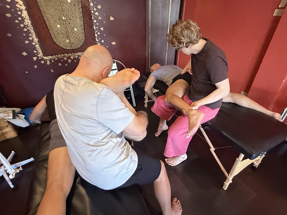
Remedial massage course
The most consequential decision a skilled therapist makes rarely involves technique. It involves timing. Two patients can present with what looks like the same hamstring injury, the same antalgic posture, the same restricted hip flexion, yet the appropriate manual response can be entirely different depending on where they sit in the biological arc of healing. Get that timing wrong and even technically flawless work can delay recovery, exacerbate pain, or destabilize a tissue that was quietly doing exactly what it needed to do.
I founded RSM International Academy because I kept encountering practitioners, skilled ones, who had learned techniques without learning the biology underneath them. They could execute deep effleurage, trigger point compression, and cross-fiber friction with precision. What they hadn't been taught was when those tools were appropriate and when they were actively harmful.
What Soft Tissue Injuries Reveal About Healing
Muscle, tendon, ligament, and fascia each carry distinct mechanical properties and distinct healing timelines. Conflating them leads to generic treatment that underserves every patient.
Muscle is highly vascularized, which means it heals faster than tendon or ligament. A grade I muscle strain typically completes its initial inflammatory phase within 72 hours. Tendons, by contrast, are hypovascular and metabolically slow. Chronic tendinopathy is a degenerative condition characterized by disorganized collagen and the near-absence of inflammatory cells. Treating it as though it were an acute inflammatory injury is a diagnostic error that no amount of technical skill compensates for.
Fascia transmits load across anatomical compartments. When an area is disrupted, the mechanical effects propagate. A runner with plantar fascia restriction may develop symptomatic hip flexor tension months later, and the foot is the last place an inexperienced therapist looks.
The Three Phases of Soft Tissue Repair: A Therapist's Clinical Map
Healing unfolds across three overlapping phases. The condition of the tissue at any given phase should dictate how manual therapy is delivered.
Inflammatory phase (days 1–5)
Vasoconstriction is followed by vasodilation; plasma fills the interstitial space; bradykinin stimulates nociceptors. This is not a problem to be suppressed. It is the environment in which phagocytes clear cellular debris and fibroblasts mobilize. Premature deep work amplifies this phase rather than shortening it. Useful manual intervention at this stage is light and circulatory: gentle effleurage proximal to the injury site to support lymphatic drainage, nothing more.
Proliferative phase (days 5 through approximately 6 weeks)
Fibroblasts populate the injured zone and synthesize Type III collagen, a fine, randomly organized matrix that lacks tensile strength. You can feel it: a soft resistance without resilience, perceptibly different from healthy muscle tissue. This phase is both an opportunity and a risk. The tissue is responsive to mechanical input, meaning targeted massage can begin to influence collagen organization. It is not yet strong enough to tolerate aggressive loading. Cross-fiber friction applied judiciously in the late proliferative stage can orient collagen fibers along lines of functional stress. Applied too early, it creates microtrauma on top of an already compromised structure.
Remodeling phase (3 weeks to 12 months)
Type III collagen is gradually replaced by the stronger, more organized Type I. Scar tissue heals three-dimensionally, reaching across fascial planes and binding structures that should glide independently. In a hamstring strain, the repaired sheath can adhere to adjacent myofascial layers, producing dull posterior thigh pain in an athlete who has otherwise returned to full training. Deep tissue massage techniques, myofascial release, and specific soft tissue mobilization during remodeling address these adhesions directly. The therapist's goal is to restore independent movement between tissue layers, not simply to reduce bulk pain.
Technique Selection for Soft Tissue Treatment: Matching the Tool to the Phase
There is no universally superior technique. There are techniques that are appropriate or inappropriate to a given tissue, at a given stage, in a given patient.
Longitudinal effleurage directed proximally is the foundational circulatory technique for subacute injuries, improving venous return and providing continuous palpatory feedback. Petrissage is appropriate for the muscle belly once the acute phase has resolved, targeting fascial extensibility rather than the injury site itself.
Deep transverse friction, applied correctly, introduces a precise mechanical force perpendicular to fiber direction, promoting collagen organization, reducing adhesion formation, and producing localized analgesia via mechanoreceptor stimulation. The conditions for appropriate application are specific: the acute inflammatory phase must have passed, the lesion must be accurately located, and the friction must reach the target tissue with sufficient depth. Applied to the wrong phase or the wrong location, it accomplishes little beyond irritation.
Sustained myofascial release addresses the tensional properties of the fascial matrix directly. Its clinical utility in the late proliferative and remodeling phases for restoring tissue glide is well-established in practice. Assisted soft tissue mobilization approaches, including instrument-assisted variants, extend the therapist's palpatory reach into deeper fascial layers. Their value lies not in aggressive scraping but in the precision with which they identify restrictions that hands alone might not detect.
The Compensation Problem in Injury Recovery
One of the most persistent clinical errors is treating the site of pain rather than the system that produced it. Injuries consistently generate compensatory movement patterns. When a muscle is inhibited by pain, adjacent structures take over its mechanical role, and those patterns often become self-perpetuating, generating a secondary layer of dysfunction that a purely local focus will never resolve.
Every patient presenting with a soft tissue injury requires a postural and movement assessment that maps the compensation pattern. The therapist who identifies that a client's recurring shoulder impingement syndrome is being driven by chronic pectoral shortening secondary to a protected rib injury from two years prior is doing something qualitatively different from the therapist who massages the rotator cuff in isolation. Technique without context is just pressure.
Palpation, Pain Science, and the Limits of Local Tissue Work
Palpation is the interface between the therapist's knowledge and the patient's biology. The ability to distinguish between a muscle belly that is dense and overactive, a fascia that is dehydrated and restricted, a tendon that is thickened and nodular versus uniformly taut, requires thousands of hours of clinical contact and an actively questioning mind. We teach palpation at RSM as a form of dialogue with the soft tissue: what is the tone of this tissue relative to its neighbors? Where is independent movement lost between layers? Is the pain sharp and superficial, or deep and referred?
Those distinctions matter because not all chronic pain follows local tissue pathology. When peripheral nociception is sustained long enough, central processing changes. The dorsal horn becomes sensitized, pain thresholds drop, and the relationship between tissue damage and pain perception becomes decoupled. A patient in a sensitized state may experience significant pain from light palpation of an area where the original injury has long since remodeled. In these cases, techniques that down-regulate the nervous system, slow broad-contact effleurage, sustained gentle compression, breathing-guided work, are clinically more productive than targeted deep tissue treatment. This is not a concession to placebo. It reflects current nociception neuroscience, and any serious sports medicine curriculum should address it directly.
Why Clinical Reasoning Matters as Much as Technical Skill
A practitioner can have excellent hands and poor outcomes if those hands are not guided by accurate clinical reasoning. Deep friction on an acutely inflamed tendon. Aggressive effleurage on a tissue in active remodeling. Trigger point compression on a sensitized patient who needed down-regulation. The techniques themselves were not wrong. The application was.
What separates a competent technician from a skilled clinician is the capacity to read the tissue, interpret what it tells you, and select the appropriate intervention from that reading rather than from habit. That capacity is built on anatomy, physiology, pathology, and supervised clinical time. It does not come from learning more techniques. It comes from understanding the ones you already have well enough to know when not to use them.
That is the orientation we build into every module of our Remedial Massage Course: anatomy and physiology as the foundation, palpatory skill developed through practice, and clinical reasoning tested against real tissue rather than an idealized protocol.
Common Orthopedic Conditions Treated with Massage: A Clinical Perspective
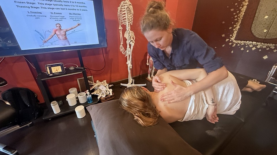
Orthopedic massage course
Most therapists encounter orthopedic pathology the hard way: a client presents with shoulder pain they've had for two years, a diagnosis that keeps shifting, and a stack of imaging that changes little except the billing codes. This is the clinical reality of orthopedic work, and it is why massage applied with genuine anatomical precision looks so different from massage applied with good intentions.
What follows is a structured look at the conditions I see most frequently in both my own practice and in the clinical work of RSM students. These are not exotic diagnoses. They are the workhorses of musculoskeletal practice, and getting them right matters.
Rotator Cuff Tendinopathy: Where Shoulder Pain Actually Lives
Few orthopedic complaints generate more therapeutic confusion than rotator cuff tendinopathy. Clinicians trained in older frameworks still reach for anti-inflammatory language, yet the tissue pathology in chronic presentations is predominantly degenerative, not inflammatory. Collagen disorganization and failed healing responses characterize the tendon. Treating a degenerative tendon as inflamed is one of the most persistent errors in shoulder pain management.
The clinical value of massage here lies in reducing hypertonicity within the rotator cuff muscles and the scapular stabilizers, particularly pectoralis minor, that alter mechanics and narrow the subacromial space. Cross-fiber techniques at the musculotendinous junctions, combined with careful glenohumeral mobility work, help restore pain-free range. The goal is not to fix the tendon through pressure; it is to modify the mechanical environment so that loading becomes tolerable again.
IT Band Syndrome and the Lateral Hip Complex
The iliotibial band does not stretch in any clinically meaningful sense. Lateral knee pain in runners is rarely caused by the band being tight; it is caused by compressive forces at the lateral femoral epicondyle, driven by altered hip mechanics and posterior hip weakness. Rolling the IT band until the athlete winces does nothing useful to the tissue.
The most productive orthopedic massage approach addresses the proximal drivers first: TFL, gluteus medius, and the lateral quadriceps. The distal knee is often the site of pain and rarely the source of dysfunction. Experienced therapists recognize this difference. It is the organizing principle that separates competent orthopedic work from symptom-chasing.
Plantar Fasciitis: Managing Load Through Orthopedic Therapy
The plantar fascia functions as a passive tension cable during the propulsive phase of gait. Dysfunction presents as morning stiffness that eases with movement before worsening under prolonged load. Like rotator cuff and patellar tendinopathy, chronic cases show degenerative tissue change, not active inflammation.
Massage directed at the intrinsic foot muscles reduces passive strain on the fascia, but the gastrocnemius-soleus complex deserves equal attention. Limited ankle dorsiflexion is one of the most consistently associated factors in plantar fascia pathology, and reducing posterior leg tightness is often more impactful than direct foot treatment. Trigger point referral from soleus into the heel frequently mimics plantar fascia pain; missing this confounds both assessment and outcomes.
Lumbar Pain Treatments and Why Tissue Work Alone Is Never Enough
Low back pain is the most common musculoskeletal complaint in adults worldwide and the most frequently overtreated with passive interventions. Massage has a legitimate role, but the therapist who treats the back without interrogating the hip, pelvis, and thoracic spine is working with a partial picture.
The multifidus exhibits atrophy and functional inhibition following disc injury or prolonged pain, maintained by altered motor programming rather than structural loss. Massage cannot rehabilitate it directly. What massage can do is reduce the superficial erector spinae overactivity that accompanies multifidus inhibition, lower the overall pain state, and improve mobility in adjacent regions creating compensatory loading. I teach RSM students to perform orthopedic assessment before any lumbar treatment, because the contraindications in this region carry genuine clinical weight.
Tennis Elbow: How Common Conditions Become Chronic
Lateral epicondylalgia presents a clinical picture that changed significantly once researchers examined the tissue directly. The extensor carpi radialis brevis tendon shows the same degenerative changes seen in rotator cuff and plantar fascia pathology. Sustained compression and eccentric load drive the condition.
Massage reduces compressive load on the ECRB insertion by addressing the surrounding extensor mass and brachioradialis. Cervical spine involvement is consistently underappreciated in chronic elbow presentations: referral from C5-C6 can sensitize the lateral elbow and surrounding soft tissue, and any therapist who treats elbow pain without assessing cervical mobility and neural tension is missing a meaningful part of the picture.
What Orthopedic Massage Actually Requires
The common thread running through every condition above is this: orthopedic massage is not a set of techniques applied to a symptomatic region. It is a clinical reasoning process that uses manual therapy as one tool within a larger understanding of tissue biology, loading mechanics, and movement dysfunction.
This is what I try to establish in RSM's Orthopedic Massage Course: not a new collection of strokes, but a systematic way of asking better questions. What tissue is involved? What stage of pathology is it in? What structures are driving the load on the symptomatic site?
A therapist who answers those questions accurately before their hands make contact will consistently outperform one who applies skilled technique without clinical direction. That framework is what separates orthopedic massage from general soft tissue work, and it is what makes the difference in the complex, chronic presentations that define any serious clinical practice.
Understanding the Clinical Benefits of Regular Fascia Massage
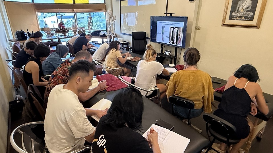
Dynamic Myofascial release course
When Jean-Claude Guimberteau first slid an endoscope under the skin of a living human subject, he did not find the neat, stratified layers depicted in 19th-century anatomy books. Instead, he captured a shimmering, chaotic, and exquisitely organized world of microvacuoles and fibrils, a sliding system that defies the static models often taught in medical school. This living matrix, the fascia, is far more than a passive biological packing material. During my years developing the curriculum at RSM International Academy, I have come to view this tissue as the primary organ of form and the fundamental mediator of human movement. For the advanced practitioner, whether a doctor, a physiotherapy specialist, or an experienced bodyworker, shifting focus from the discrete muscle unit to the continuity of the myofascial web is a necessary evolution in clinical efficacy.
The historical tendency to dissect the body into independent parts has served our understanding of discrete mechanics but has largely failed to explain the systemic nature of chronic pain and postural collapse. When we engage in myofascial release massage, we are not simply rubbing a sore spot. We are communicating with a complex, fluid-filled network that responds to mechanical load through a process known as mechanotransduction. This biological signaling converts the physical pressure of a massage into biochemical signals, stimulating the remodeling of the extracellular matrix. By addressing the body as a single, uninterrupted unit of connective tissue, we move away from symptomatic treatment toward a structural logic that addresses the root of dysfunction.
Biotensegrity and the Dynamics of Fascia
The conceptual shift toward understanding myofascial continuity is grounded in the principle of biotensegrity. In this model, the skeleton is not a weight-bearing frame like the studs of a house, but rather a series of compression-resistant struts suspended within a continuous sea of tension provided by the fascia. When this tension is balanced, the body moves with an effortless efficiency. However, when fascial restrictions develop due to trauma, repetitive strain, or sedentary behavior, the entire system must compensate. A restriction in the plantar fascia can manifest as a cervical spine issue, as the body attempts to maintain a level horizon through the kinetic chain.
Regular manual manipulation of these structures ensures that the sliding surfaces of the body remain hydrated and mobile. At the microscopic level, the tissue is lubricated by hyaluronan, a glycosaminoglycan that functions as a molecular lubricant. When the body remains static, this substance can become gel-like and viscous, leading to densification and the sensation of stiffness. The heat and specific shear forces applied during a myofascial massage encourage a thixotropic effect, transitioning this lubricant back into a fluid state. This fluid exchange is vital for cellular health, as the fascia acts as the delivery system for nutrients and the drainage system for metabolic waste.
Mitigating Chronic Pain via Myofascial Release
The neurological impact of a sustained release is perhaps its most profound attribute. We must recognize that the fascia is our richest sensory organ, packed with six times as many sensory nerve endings as the muscles themselves. These include Ruffini endings, which respond to slow, lateral pressure, and Pacini corpuscles, which detect rapid changes in pressure and vibration. When we provide a targeted myofascial release massage, we are essentially recalibrating the nervous system's perception of safety and threat.
In cases of fibromyalgia or long-term structural trauma, the nervous system often becomes sensitized, maintaining a state of high tension even when no immediate threat is present. This fascial armor creates a feedback loop where pain leads to further restriction, and restriction leads to more pain. By applying a slow, deliberate myofascial approach, we can down-regulate the sympathetic nervous system. The result is a significant reduction in chronic pain that often persists long after the session has ended, as the tissue begins to adopt a more functional, less defensive posture.
Neurological Feedback in Myofascial Massage
Professional practitioners understand that the goal of massage therapy in a clinical context is to break these self-perpetuating cycles. We are looking for the melt, that specific moment when the fascial tissues yield under the hand. This is not a product of force, but of time and presence. Because the fascia is viscoelastic, it does not respond well to sudden, aggressive movements. Instead, it requires a sustained load to reach the creep phase, where the fibers actually begin to lengthen and reorganize.
- Golgi Tendon Organs: Located at the junctions of muscles and tendons, these respond to muscular contraction and deep pressure, helping to reset muscle tone.
- Ruffini Endings: These are sensitive to tangential forces and sustained lateral stretch, making them the primary targets during structural integration.
- Pacini Corpuscles: These receptors respond to rapid pressure changes and are vital for proprioception.
- Interstitial Receptors: These are linked to the autonomic nervous system, influencing heart rate and blood pressure.
By engaging these receptors through skilled tissue massage, we do more than just provide pain relief. We are enhancing the body's internal mapping system. An athlete with a well-hydrated, mobile fascial system has superior proprioception, allowing for faster reactions and a lower risk of injury. This is a primary reason why rehabilitation protocols in elite sports are increasingly focusing on the connective matrix rather than just the contractile muscle fibers.
Tissue Plasticity and Pain Relief
For the professional athlete or the high-performance mover, the recovery phase is as critical as the training phase. When we apply deep tissue techniques within a sports medicine framework, we are facilitating the rapid removal of post-exercise inflammatory markers. However, we must distinguish between standard deep tissue massage and the specific structural work we teach at RSM. I have often explained to my students that improved movement is the natural byproduct of a body that no longer has to fight itself. When the fascia is constricted, the muscles must work twice as hard to achieve the same result.
Regular massage therapy targeting the connective chains ensures that the recoil property of the fascia is preserved. This elastic recoil is what allows a runner to spring off the pavement or a golfer to generate power through the torso. Without it, we are relying purely on muscular effort, which is metabolically expensive and mechanically inefficient. The application of deep tissue massage in this context serves to hydrate the tissue and break down adhesions that form in the superficial and deep layers.
Professional Massage and Structural Rehabilitation
In the clinical setting, we often encounter patients who have sought relief through various pharmaceutical or surgical interventions with limited success. This is frequently because the source of the pain is not in the bones or the nerves, but in the distortions of the connective matrix. Addressing these through regular massage provides a non-invasive pathway to systemic health. The benefits extend beyond the physical, as the fascia is intimately connected to the autonomic nervous system.
Our Myofascial Release Course is designed for the intelligent professional who recognizes that the old models of bodywork are no longer sufficient. Whether you are dealing with the complex presentation of fibromyalgia, the repetitive strain of a professional athlete, or the chronic postural issues of the modern office worker, the ability to work effectively with the fascia is a transformative skill.
Through regular massage, we encourage the lymphatic drainage necessary to clear inflammatory cytokines, effectively cleaning the internal environment of the body. The commitment to regular massage therapy is an investment in the longevity of the human frame. As we age, our fascia naturally tends to lose its water-binding capacity and becomes more brittle. By consistently applying the principles of myofascial work, we can slow this process significantly, maintaining the fluidity and grace of movement that characterizes youth. At RSM, we emphasize that the therapist is a facilitator of the body's own healing intelligence. We are not fixing the client; we are removing the fascial restrictions that prevent the body from fixing itself. This perspective requires humility and technical proficiency, as the fascia is the key to a new paradigm of health.
